


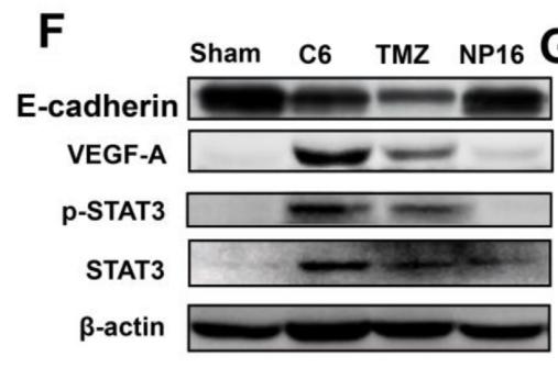
. Cell migration and invasion of A2780 (a, b) and HEY cells (c, d) were detected via Transwell analysis, and the expression of GTP-RhoA, total RhoA, β-catenin, VEGF, MMP2, and MMP7 in A2780 and HEY cells was detected via western blotting (e–g).")
 The expression of VEGF mRNA was determined by qPCR at days 1 and 3. (B) The expression of CD31 mRNA. (C) The expression of EMCN mRNA. (D–G) The expression of VEGF, EMCN and CD31 proteins were determined by Western blot analysis for 3 and 7 days. All the data were confirmed by three repeated tests. Data were mean ± S.D. *p < 0.05 vs. the control group at the same day.#p < 0.05 vs. the control group at the same day")
 and the typical phenotype of adipocytes (stained with Oil Red O). E, Flow cytometry showed the ADSCs
expressed more stem cell markers (CD90, CD44a and CD29), but less haematopoietic and endothelial markers (CD34, CD11b and CD45).
F, Immunofluorescence analysis of the expression of RFP in transfected ADSCs. G,H, Western blot analysis showed markedly increased
the levels of VEGF in transfected ADSCs. I, Immunofluorescence analysis of the expression of GFP in transfected ADSCs. J,K, Western
blot analysis showed increased the levels of GDNF in transfected ADSCs. L, Immunofluorescence of ADSCs that co-expressed RFP and
GFP. M,N, Western blot analysis of ADSCs co-transfected with VEGF and GDNF. All values are represented as the mean ± SD from three
independent experiments, each with three replicates. Statistically significant differences from the control group are denoted as follows:
***P < .001, **P < .01 and *P < .05 (independent samples t test)")
 Confocal images of U87 cells treated with Exos-Dox and PC-Exos-Dox. Green: exosomes
labeled by FITC. Red: Dox fluorescence. Blue: nuclei stained by DAPI. Scale bar, 20 µm. B) Cellular uptake of Exo-Dox and PC-Exos-Dox by U87 cells
as assessed by flow cytometry, Dox signal detection. C) Cell viability of U87 cells exposed to different concentrations of Exos-Dox and PC-Exos-Dox.
D) Confocal images of U87 cells treated with Exos-anti-miR21 and PC-Exos-anti-miR21. Green: exosomes labeled by FITC. Red: Cy5 fluorescence of
anti-miR21. Blue: nuclei stained by DAPI. Scale bar, 20 µm. E) Cellular uptake of Exo-anti-miR21 and PC-Exos-anti-miR21 by U87 cells as assessed by
flow cytometry, Cy5-anti-miR21 signal detection. F) Western blot analysis of protein expression in U87 cells 48 h after treatment. GAPDH was used as a
loading control. Data in (B), (C), and (E) are presented as the mean ± SD (n = 3). The significance levels are shown as **p < 0.01.")
 The effects of HMGB1/sRAGE on RCC-stimulated VEGF and VEGFR2 proteins expression in HUVEC were detected by Western blot analysis. *P,0.05, **P,0.01 vs Con-siRNA group; ##P,0.01 vs Con-Vec group; ^^P,0.01 vs sRAGE group.")
 in AA patients with KB (n = 3). A, Relative mRNA expression. B, Protein brands. C, Relative protein expression.Normal, normal bone marrow tissues (n = 5); AA, bone marrow tissues of AA patients with KB (n = 10). **P < 0.01 vs Normal;*P < 0.05 vs normal. AA, aplastic anemia; KB, kidney‐deficiency and blood‐stasis; VEGF, vascular endothelial growth factor; VEGFR, VEGF receptor")

 The bands of protein
expression. (B)-(F) Correlation analysis between the depression-like behaviors and protein expression. Statistical analyses of single comparison were conducted
through the Student’s t-test. All data are presented as mean ± SD, *p < 0.05, **p < 0.01, n = 3 per group.")

. Magnification, x400; scale bar, 50 µm. **P<0.01 vs. the AS‑IV group; ##P<0.01 vs. the AS‑IV‑CID755673 group; $P<0.05 vs. the Model group; &P<0.05 vs. the CID755673 group. AS‑IV, astragaloside IV; PKD1, protein kinase D1; HDAC5, class IIa histone deacetylase 5; VEGF, vascular endothelial growth factor.")

 Immunohistochemical staining: The immunoreactive cells were stained yellow and observed in the spinal cord. Typical immunoreactive cells are marked by arrows. Few immunoreactive cells were seen in the sham group, and the number of immunoreactive cells was higher in the SCIC group than in the SCIO group. Scale bars: 500 μm. (B) Percentage of immunoreactive cells in the injured spinal cord: The percentages of HIF-1α- and VEGF-immunoreactive cells were higher in the SCIC group than in the SCIO group (*P < 0.05; mean ± SD, n = 8; one-way analysis of variance followed by the Bonferroni correction in various groups). HIF-1α: Hypoxia-inducible factor 1α; SCIC: spinal cord injury with closed canal; SCIO: spinal cord injury with open canal; VEGF: vascular endothelial growth factor.")
 Western blot bands: The bands were more obvious in the SCIC group than in the SCIO group. (B) Percentage of objective protein expression in the injured spinal cord. The expression of HIF-1α and VEGF proteins was higher in the SCIC group than in the SCIO group (*P < 0.05; mean ± SD, n = 8; one-way analysis of variance followed by the Bonferroni correction in various groups). HIF-1α: Hypoxia-inducible factor 1α; SCIC: spinal cord injury with closed canal; SCIO: spinal cord injury with open canal; VEGF: vascular endothelial growth factor.")
, and then gradually decreased. GAPDH, glyceraldehyde-3-phosphate dehydrogenase; HUVEC, human umbilical
vein endothelial cell; VEGF, vascular endothelial growth factor.")
 Immunohistochemistry was used to detect the expression of vascular endothelial growth factor (VEGF) in the flaps of the three groups of rats; (B) VEGF
contents in the flaps of the three groups of rats were expressed by integral absorbance (IA) value. **P < 0.01, CDPC-L group vs. control group; **P < 0.01, CDPC-L
group vs. CDPC-H group.")
 Immunohistochemistry was used to
detect the expression of vascular endothelial
growth factor (VEGF) in the flaps of the three
groups of rats; (B) VEGF contents in the flaps of
the three groups of rats were expressed by integral absorbance (IA). **P < 0.01, DEX-L
group vs. control group; **P < 0.01, DEX-L
group vs. DEX-H group. n = 6 per group.")

 the Immunohistochemical evaluation of VEGF, Notch1, and Dll4 in the zone 2 of the two groups. The
immunohistochemistry test and observed under original magnification 400 (b) Integral absorbance (IA) values were detected to
compare the level of VEGF, Notch1, and Dll4.")
 Expression of VEGF, Notch1, and Dll4 by
Western blot. (b) Densitometry results of VEGF, Notch1, and
Dll4 proteins expression (p < 0.01 vs. the control group).")
 and the typical phenotype of adipocytes (stained with Oil Red O). E, Flow cytometry showed the ADSCs expressed more stem cell markers (CD90, CD44a and CD29), but less haematopoietic and endothelial markers (CD34, CD11b and CD45). F, Immunofluorescence analysis of the expression of RFP in transfected ADSCs. G,H, Western blot analysis showed markedly increased the levels of VEGF in transfected ADSCs. I, Immunofluorescence analysis of the expression of GFP in transfected ADSCs. J,K, Western blot analysis showed increased the levels of GDNF in transfected ADSCs. L, Immunofluorescence of ADSCs that co‐expressed RFP and GFP. M,N, Western blot analysis of ADSCs co‐transfected with VEGF and GDNF. All values are represented as the mean ± SD from three independent experiments, each with three replicates. Statistically significant differences from the control group are denoted as follows: ***P < .001, **P < .01 and *P < .05 (independent samples t test)")
 in a penile mid‐shaft specimen 2 wk after BCNI and treatment. F, Quantitative analysis of the GDNF‐positive area. G,H, Representative immunofluorescence of VEGF in a penile mid‐shaft specimen, and quantitative analysis of the VEGF immunofluorescence‐positive area. Data are depicted as the mean ± SD from n = 6 animals per group (*P < .05). Immunofluorescence was analysed using one‐way ANOVA followed by the S‐N‐K test. Quantitative analysis of immunohistochemistry was performed using the Kruskal‐Wallis H test")
 in a penile mid‐shaft specimen 2 wk after BCNI and treatment. F, Quantitative analysis of the GDNF‐positive area. G,H, Representative immunofluorescence of VEGF in a penile mid‐shaft specimen, and quantitative analysis of the VEGF immunofluorescence‐positive area. Data are depicted as the mean ± SD from n = 6 animals per group (*P < .05). Immunofluorescence was analysed using one‐way ANOVA followed by the S‐N‐K test. Quantitative analysis of immunohistochemistry was performed using the Kruskal‐Wallis H test")
 Detection of HMEC-1 cell proliferation by a CCK-8 assay. (B) Images of HMEC-1 cells in scratch
sites. Scale bar, 250 mm. (C) Scratch length statistics of three group. (D) Images of angiogenesis length and the number of branch points at 4 and 8 h in HMEC-1 cells. (E) Detection of
p-ERK1/2, ERK1/2, and VEGFA in HMEC-1 cells by western blotting. (F) Bar graph of gray values of p-ERK1/2, ERK1/2, and VEGFA protein bands. HMEC-1: HMEC-1 cultured alone.
Exosome þ HMEC-1: HMEC-1 co-cultured with untreated exosomes. Elongation-exosome þ HMEC-1: HMEC-1 co-cultured with exosomes subjected to mechanical stress treatment.
*p < 0.05, **p < 0.01 vs. HMEC-1. #p < 0.05, ##p < 0.01vs. Exosome þ HMEC-1.")

Quantitative reverse-transcription polymerase
chain reaction of Vegf, Alp, Osx, Ocn, Trap, and
Ctsk in the calluses of sham-operated groups
and osteotomized condylar necks of control
(CON) and conditional knock-out mice (CKO)
on POD14 and 28. Data reflect mean ± SEM. (b)
Immunofluorescence of VEGF on POD14 and
VEGF and CD31 on POD28 in the calluses. Scale
bars =100 μm. (c) ALP of calluses on POD28 in
control and cko mice. The green arrows show
positively stained cells. The area of higher
magnification shows immunofluorescence of
OSX. Lower magnification, scale bars =200 μm;
higher magnification, scale bars =100 μm. (d)
TRAP-staining in control and cko groups on
POD28. The red arrows show cells positive for
TRAP expression. The area of higher magnification shows immunofluorescence of CTSK.
Lower magnification, scale bars =200 μm;
higher magnification, scale bars =100 μm.
ns ≥ 0.05 (no significance), *p < 0.05,
**p < 0.01, ***p < 0.001, ****p < 0.0001. (For
interpretation of the references to colour in this
figure legend, the reader is referred to the web
version of this article).")
 tubulin dynamics, (B) angiogenesis and (C) cell cycle. CK: Control check; S: Single; P: Polymer.")
. E, G. The effect of exercise on the expression of VEGFC in lung cancer tissues (n=6). Note: &, && respectively represent a significant difference compared with the Saline group (P<0.05),(P<0.01).")
 The overexpression efficiency of circDIP2C was determined by qRT-PCR in HUVECs. (B-H)
HUVECs were singly treated with ox-LDL, ox-LDL + Vector and ox-LDL + circDIP2C, and untreated HUVECs served as a Control. (B) Cell viability was determined by
CCK-8 assay. (C) Tube formation was detected by tube formation assay. (D and E) The levels of ROS and MDA were measured by ROS and MDA determination assays.
(F and G) SOD and LDH activity were determined by SOD and LDH activity assay, respectively. (H) Western blot analysis was employed to detect the protein
expression of NLRP3, pro-Caspase 1, Caspase 1, pro-IL-1β, IL-1β and VEGF. *P < 0.05.")
 IHC staining was carried out to delineate the expression of PCNA protein in Skov3 cells. Real-time PCR was employed to detect the transcriptional levels of (B) P53 and (C) P21. (C, D, E, and F) Western blot analysis was conducted to measure key proteins of several signaling pathways. *, p<0.05 vs control group, * *, p<0.01 vs control group")
 The number of total cells, macrophages and neutrophils in the BALF was counted
with a hemocytometer. (B) Effect of loganin on lung MPO activity. (C) Relative protein levels of VEGF in the lung tissues by western blotting. (D) Immunohistochemistry for F4/80 in the lung tissues. Bars = 50 μm. Data were shown as means ± SD. n = 6. *p < 0.05 vs. sham. #p < 0.05 vs. CLP. ALI, acute lung injury; BALF,
bronchoalveolar lavage fluid; MPO, myeloperoxidase; VEGF, vascular endothelial growth factor; CLP, cecal ligation and puncture.")
 Immunofluorescence staining of blood vessels (green) in the hippocampus on the 14th day after grafting. Scale bar = 100 μm. (b) Quantification of vascular density in the 2VO+Cell and 2VO+Cell+EA groups. (c) Representative Western blots of VEGF on the 14th day after grafting. (d) The densitometric analysis of VEGF level detected from the hippocampus in each group (one-way ANOVA, F = 13.574, P < 0.001). Values are mean ± SEM (N = 5 rats/per group). ∗P < 0.05 and ∗∗∗P < 0.001.")
 and protein (d) expression in naive T cells, Caki-1, and 786-O cells treated with hucMSC-Exo detected using RT-qPCR and Western blotting. e Binding relation between miR-182 and VEGFA verified by dual-luciferase reporter assay. f sequence information of miR-182 in mice and humans predicted using miRBase website. g the distribution of miR-182 in MSCs predicted using RNAlocate website. Cell experiment is repeated 3 times. Data are shown as mean ± standard deviation. **p < 0.01 vs. PBS group")

: representative immunoreactive bands of ODC, VEGF, MMP-9, GAPDH, total and phosphorylated Erk1/2 using specific antibody. (B–E): representative quantification of ODC, phosphorylated Erk1/2, VEGF, MMP-9. (F): determination of MMP-9 activity. * p < 0.05, ** p < 0.01, *** p < 0.001 compared with MCF-7/ADR cells, ## p < 0.01, ### p < 0.001, comparison between MCF-7/ADR group and MCF-7 group.")
 mRNA expression levels of osteogenesis-related markers in BMSCs following treatment with/without melatonin. (B) Protein expression levels of osteogenesis-related marker VEGF. (C) VEGF content secreted in the supernatant liquid assessed by ELISA kits. (D) Immunofluorescent images of BMSCs stained for VEGF (scale bars, 100 μm). All the experiments were repeated at least 3 times independently. BMSCs, bone marrow mesenchymal stem cells; VEGF, vascular endothelial growth factor. The data are presented as means ± SEM. * p < 0.05, ** p < 0.01 vs. control group, # p < 0.05, ## p < 0.01 vs. 100 nM melatonin group.")
 mRNA expression levels of osteogenesis-related markers in BMSCs following treatment with/without melatonin. (B) Protein expression levels of osteogenesis-related marker VEGF. (C) VEGF content secreted in the supernatant liquid assessed by ELISA kits. (D) Immunofluorescent images of BMSCs stained for VEGF (scale bars, 100 μm). All the experiments were repeated at least 3 times independently. BMSCs, bone marrow mesenchymal stem cells; VEGF, vascular endothelial growth factor. The data are presented as means ± SEM. * p < 0.05, ** p < 0.01 vs. control group, # p < 0.05, ## p < 0.01 vs. 100 nM melatonin group.")

 The MAPK14 3′-UTR containing the wildtype or mutant miR-200a-3p binding sequence was inserted into downstream of the luciferase reporter vector. The mutated sequences are italicized. (B) The dual luciferase reporter assay revealed that the luciferase activity controlled by MAPK14 3′-UTR was inhibited by ectopic miR-200a-3p expression in 293T cells. (C) miR-200a-3p was highly expressed or knocked down in HUVEC by lipofectamine 2000 transfection. QRT-PCR analysis was performed to measure the expression levels of miR-200a-3p in HUVEC after treatment with miR-200a-3p mimic, mimic control or miR-200a-3p inhibitor, inhibitor control. (D) The mRNA levels of MAPK14, c-jun, ET-1 and VEGFA were determined by qRT-PCR in HUVEC transfected with miR-200a-3p mimic, mimic control or miR-200a-3p inhibitor, inhibitor control. (E) Western blot analysis of p38, phosphorylated p38, c-jun, phosphorylated c-Jun, ET-1 and VEGFA protein levels in HUVEC transfected with miR-200a-3p mimic, mimic control or miR-200a-3p inhibitor, inhibitor control. (F) Quantification of protein results in panel E. Data were expressed as mean ± SEM. Between group differences were assessed by the student’s t test, respectively. *p < 0.05, **p < 0.01, ns, not significant.")
 HIF-1 and VEGFA expression in fraxetin-treated PANC-1 and Patu8988 cells as seen on a Western blot. (B) Western blot analysis of HIF-1 and VEGFA expression in fraxetin-treated animal xenograft models. (C) IHC staining for HIF-1α in fraxetin-treated models. Bar = 100 μm. (D–G) Glucose metabolism assay shows downregulated oxygen consumption rate (OCR), basal respiration, spare respiration, maximal respiration, and ATP production in fraxetin-treated PANC-1 and Patu8988 cells. (H–K) Glucose metabolism assay showing reduced levels of extracellular acidification rate (ECAR), basal glycolysis and compensatory glycolysis in fraxetin-treated PANC-1 and Patu8988 cells. (L) GLUT1 expression in fraxetin-treated PANC-1 and Patu8988 cells as seen on a Western blot. Data were presented as the mean ± standard deviation, and were analyzed by One-way ANOVA with Bonferroni’s post-hoc test and two-sided Student’s t-test. *P < 0.05; **P < 0.01, ***P < 0.001.")
 Laser Doppler images show blood perfusion in the flap (scale bar: 1 cm) The H&E staining between three groups showing the vessels (scale bar: 50 and 25 μm) (B) Signal intensity of blood flow in skin flaps among the three groups (C) The representative fluorescence image of VEGF with DAPI (nuclei) (scale bar: 25 μm) (D) Mean vessel density of mice skin flap tissue (E) The percentage of VEGF positive cells in dermal layer of skin flaps. All data represent mean ± SD. **p ≤ 0.01 compared to the control group.")
 Immunohistochemistry images of VEGF. All images were obtained at identical magnification, ×200, scale bar = 50 μm. (B) Quantitative analysis of VEGF content (n = 3). Data are represented as mean ± SEM. **P")
 Hif-1α and VEGF expression was assessed by RT-qPCR in CMVECs. (B) Hif-1α and VEGF expression was assessed by western blot in CMVECs before and after AngII treatment. (C) Hif-1α and VEGF expression was assessed by RT-qPCR in AngII-induced CMVECs after treatment with normal saline, EUE, or EUE + WR-1065. (D) Hif-1α and VEGF expression was assessed by western blot in AngII-induced CMVECs after treatment with normal saline, EUE, or EUE + WR-1065. Data were displayed as mean ± standard deviation, N = 3. *P < 0.05 compared with the control or saline group; # P < 0.05 compared with the EUE group. AngII, angiotensin II; CMVEC, cardiac microvascular endothelial cell; EUE, Eucommia ulmoides extract; Hif-1α, hypoxia-inducible factor-1α; VEGF, vascular endothelial growth factor.")
 Magnification after immunohistochemical staining of hypoxia‐inducible factor‐1α (HIF‐1α) and vascular endothelial growth factor (VEGF) on day 7. (B) Expression of HIF‐1α on day 7. Roxadustat (RXD) improved the expression of HIF‐1α. (C) Expression of VEGF on day 7. RXD improved the expression of VEGF. **P")
 operating at 400× magnification (a). The integral absorbances of flap VEGF, TLR4, NF-kB, IL-1β, IL-6, and TNF-α levels in the control and treatment groups (b). **P")
 Functions and pathways negatively enriched for ImP treated cells and positively enriched for S1P treated cells were shown. The differentially expressed proteins was identified and analyzed by GO and KEGG pathway analysis. (C) Protein-protein interaction network analysis of ImP down-regulating protein by STRING. (D and E) The effects of S1P and ImP on the expression levels of VEGF, HIF-1α, CD31, and Ki67 proteins in HUVEC cells detected by Western blot. (F and G) The effect of ImP and S1P on the protein expression levels of MMP2, MMP9, Ki67 and Vimentin in NIH3T3cells under coculture conditions. (H) The effect of ImP on the cell membrane localization of RhoA. ImP-L:500 nM, ImP-H: 1μΜ. Data are expressed as the mean ± SD (∗p < 0.05, ∗∗p < 0.01). The number of sample replicates for all experiments was 3 (n = 3).")
 The effect of hypoxia treatment for 4 weeks on Cul5/TRAF6/NF-κB/HIF-1α/VEGF pathway expression in lung tissues of mice, and reverse effect of pevonedistat. (a) Western blot analysis of TRAF6/NF-κB/HIF-1α/VEGF pathway expression in N4W and H4W group and statistical analysis of the proteins relative expression normalized to GAPDH. (b) Western blot analysis of TRAF6/NF-κB/HIF-1α/VEGF pathway expression in N4W + DMSO, N4W + pevonedistat, H4W+ DMSO, and H4W + pevonedistat group, and statistical analysis of the proteins relative expression normalized to GAPDH. (B) The effect of hypoxia and/or Cul 5 treatment on TRAF6/NF-κB/HIF-1α/VEGF pathway expression in PAECs. Western blot analysis of TRAF6/NF-κB/HIF-1α/VEGF pathway expression in endothelial cells exposed to normoxia and hypoxia with or without Cul 5, and statistical analysis of the proteins relative expression normalized to GAPDH. ∗p < 0.05, ∗∗p < 0.01, ∗∗∗p < 0.001. Student’s t test was used for statistical analysis in Figure 7A(a). Data are represented as the means ± SD, one-way ANOVA was used for statistical analysis in Figure 7A(b) and 7B.")
 Picrosirius red staining of the dorsal skin wound site in mice observed using polarized light microscopy after NnNV injection and 7-day treatment with PCA (green) or DHB (yellow) (n = 4). (A) Scale bar, 50 μm. (B) Quantitative data showing the percentage of type I collagen fibers in the wound bed field for each skin tissue group. (C) Quantitative data showing the percentage of type III collagen fibers in the wound bed field for each skin tissue group. (D) Proportion of type III/type I collagen fibers in the wound bed domain. (E) Immunohistochemical staining showing the expression of vascular endothelial growth factor (VEGF) and basic fibroblast growth factor (bFGF) at the margins of dorsal skin wounds in mice 7 days after NnNV injection and treatment with PCA (green) or DHB (yellow). Scale bar, 100 μm. Blank groups received injections of an equal volume of PBS buffer (PBS, black). The NnNV control group received only NnNV injection without cream application (NnNV, blue), while the Cream control group received NnNV injection with cream application but without drug loading (Cream, grey). Data are mean ± s.e.m. n = biological replicates.")
, the immunohistochemical results of Piezo1, RUNX2, BMP2, CD31, HIF-a, and VEGF are presented. Additionally, (B–G) display the corresponding quantitative immunohistochemical results. In comparison to the model group, both the blank and WBVT groups exhibited significantly higher expression levels of Piezo1, RUNX2, BMP2, CD31, HIF-a, and VEGF. These differences were statistically significant (p < 0.05).")
 and protein (B) expression in rats. Mean ± SD. n = 3. ##P < 0.01 vs sham group; **P < 0.01 vs model group.")
 Western bolt showed protein expression levels in different experimental groups.Mean ± SD represents all the statistical data. The significance levels are denoted as follows: (*p < 0.05, **p < 0.01, ***p < 0.001, ****p < 0.0001, ns indicates that the difference is not statistically significant)")
 Western blot showed the protein level of Aβ42, p-tau, S100β, NSE, APJ, and VEGF change with different treatments. (B–G) Quantitative analysis of A, protein levels of Aβ42 in LPS, 20 μmol/L mangiferin with LPS, 40 μmol/L mangiferin with LPS compared to control were 2.10, 1.67, and 1.30 times, respectively, all ***P < 0.001. One-way ANOVA, F = 111.7, ***P < 0.001. Protein levels of p-tau in LPS, 20 μmol/L mangiferin with LPS, 40 μmol/L mangiferin with LPS compared to the control are 2.60, 1.94, and 1.53 times, respectively, all ***P < 0.001. One-way ANOVA, F = 285.7, ***P < 0.001. Protein levels of S100β in LPS, 20 μmol/L mangiferin with LPS, and 40 μmol/L mangiferin with LPS compared to the control are 2.11, 1.61, and 1.38 times, respectively, **P = 0.0026 for 20 μmol/L mangiferin with LPS vs. 40 μmol/L mangiferin with LPS. One-way ANOVA, F = 241.0, ***P < 0.001. Protein levels of NSE in LPS, 20 μmol/L mangiferin with LPS, 40 μM mangiferin with LPS compared to the control are 3.57, 2.27, and 1.67 times, respectively, all ***P < 0.001. One-way ANOVA, F = 990.9, ***P < 0.001. Protein levels of APJ in LPS, 20 μmol/L mangiferin with LPS, 40 μmol/L mangiferin with LPS compared to control are 0.45, 0.63, and 0.82 in fold, respectively, **P = 0.0028 for LPS vs. 20 μmol/L mangiferin with LPS, **P = 0.0015 for 20 μmol/L mangiferin with LPS vs. 40 μmol/L mangiferin with LPS. One-way ANOVA, F = 108.3, ***P < 0.001. Protein levels of VEGF in LPS, 20 μmol/L mangiferin with LPS, 40 μM mangiferin with LPS compared to control were 0.43, 0.57, and 0.79 times, respectively, all ***P < 0.001. One-way ANOVA, F = 467.7, ***P < 0.001.")

 in acid-induced ulcerated tissues. C Statistical analysis of CD31 and VEGF (D) expression in each group. E mRNA expression levels of CD31 and VEGF (F) in each group. G Protein expression levels of CD31 and VEGF detected by western blot (WB) in each group. H The average protein expression levels of CD31 and VEGF (I). In all experiments, data were presented as mean ± SD for n = 3 biological replicates.")

 (scale bar: 100 μm). F The optical density values of HIF-1α, HSP90 and VEGF in the flaps of the three groups. G, H The intergral absorbance of HIF-1α and VEGF in the flaps of the three groups. I Western blotting of HO-1 and SOD1 expressions in the flaps of the three groups. J The optical density values of HO-1 and SOD1 in the flaps of the three groups. K Mean SOD activity of the flaps in the three groups. L Mean MDA content of the flaps in the three groups. M The optical density values of IL-6 in the flaps of the three groups. N, O The intergral absorbance of IL-6 and TNF-α in the flaps of the three groups. P Western blotting of IL-6 expressions in the flaps of the three groups. Q Immunohistochemistry for IL-6 and TNF-α expression of the flaps in the three groups (original magnification × 200) (scale bar: 100 μm). Data are presented as mean ± SD, n = 6 per group. ns P > 0.05, **P")
Schematic diagram of cells that promote angiogenesis. (B)Representative microscopic images of HUVEC after the cells were co-cultured with different macrophage cultures for 12 h. Scale bars = 250 μm. (C)Representative Transwell migration images were obtained after macrophage culture medium was added to the lower chamber alone and cultured for 24 h. Scale bars = 200 μm. (D)Representative immunofluorescence images of VEGF (green) and nucleus (blue) in HUVEC after 7 days of co-culture of cells with different macrophage media. Scale bars = 50 μm. (E) Representative images of blood vessel formation in the CAM test with the presence of scaffolds. (F)After the cells were co-cultured with different macrophage media, Representative fluorescence images and quantitative analysis of HUVEC tube formation. Scale bars = 200 μm. (G)Relative mRNA expression levels of angiogenesis related genes, including VEGF and Ang-1, in HUVEC after 7 days of co-culture of cells with different macrophage media.Data are expressed as mean ± SD (n = 3). ∗P < 0.05 and ∗∗P < 0.01 indicated significant difference compared with CON group.")
 Western blot analysis was conducted to assess VEGFA and VEGFR2 protein expression at 3, 7, 14, and 28 days post-chronic cerebral hypoperfusion. (A) Protein bands for VEGFA and VEGFR2 are displayed. (B-C) Corresponding bar graphs represent the optical density values for VEGFA and VEGFR2 protein bands in each experimental group. (D) Bar graphs present the ELISA data analysis for VEGFA protein levels across the same time points. Each group comprised n = 5 samples. * Indicates P")
 Hippo pathway related proteins expression was determined by Western blot and qRT-PCR. n = 3; (D) Cell viability assay; (E) Optical microscope images of MCMECs morphological changes; (F) Cell damage assay; (G) TUNEL assay to assess cell apoptosis rate in vitro. n = 3; (H) Transmission electron microscope images of MCMECs; (I,K) Caspase-9, Bcl-2, VEGF, FGF2 protein expression was determined by Western blot. n = 3; (J) Caspase-9, Bcl-2, VEGF, FGF2 mRNA level was assessed by qRT-PCR. n = 3.")
 H&E staining of IPF lung tissue and normal control lung tissue, showing images at 200× and 400× magnification. (B) Representative immunofluorescence images of CDKN2A (green), SOX2 (red), FOXO3 (yellow), and VEGFA (pink) expression in IPF lung tissue and normal control lung tissue (magnification: 400×). DAPI, 4′,6-diamidino-2-phenylindole. (C) Quantitative histogram of relative fluorescence intensity (n=3), with the y-axis representing fluorescence intensity relative to the healthy control group.")
| Product: | VEGFA Antibody |
| Catalog: | AF5131 |
| Description: | Rabbit polyclonal antibody to VEGFA |
| Application: | WB IHC IF/ICC |
| Cited expt.: | WB, IHC, IF/ICC |
| Reactivity: | Human, Mouse, Rat |
| Prediction: | Pig, Bovine, Horse, Sheep, Rabbit, Dog |
| Mol.Wt.: | 16-20kDa,25-30kDa,40-45kDa(Observed); 27kD(Calculated). |
| Uniprot: | P15692 |
| RRID: | AB_2837617 |
Control Products

Related Downloads
Protocols
Product Info
*The optimal dilutions should be determined by the end user. For optimal experimental results, antibody reuse is not recommended.
*Tips:
WB: For western blot detection of denatured protein samples. IHC: For immunohistochemical detection of paraffin sections (IHC-p) or frozen sections (IHC-f) of tissue samples. IF/ICC: For immunofluorescence detection of cell samples. ELISA(peptide): For ELISA detection of antigenic peptide.
Cite Format: Affinity Biosciences Cat# AF5131, RRID:AB_2837617.
Fold/Unfold
Folliculostellate cell-derived growth factor; Glioma-derived endothelial cell mitogen; MGC70609; MVCD1; Vascular endothelial growth factor A; vascular endothelial growth factor A121; vascular endothelial growth factor A165; vascular endothelial growth factor; Vascular permeability factor; VEGF A; Vegf; VEGF-A; VEGF120; Vegfa; VEGFA_HUMAN; VPF;
Immunogens
A synthesized peptide derived from human VEGFA, corresponding to a region within the internal amino acids.
Isoform VEGF189, isoform VEGF165 and isoform VEGF121 are widely expressed. Isoform VEGF206 and isoform VEGF145 are not widely expressed. A higher level expression seen in pituitary tumors as compared to the pituitary gland.
- P15692 VEGFA_HUMAN:
- Protein BLAST With
- NCBI/
- ExPASy/
- Uniprot
MNFLLSWVHWSLALLLYLHHAKWSQAAPMAEGGGQNHHEVVKFMDVYQRSYCHPIETLVDIFQEYPDEIEYIFKPSCVPLMRCGGCCNDEGLECVPTEESNITMQIMRIKPHQGQHIGEMSFLQHNKCECRPKKDRARQEKKSVRGKGKGQKRKRKKSRYKSWSVYVGARCCLMPWSLPGPHPCGPCSERRKHLFVQDPQTCKCSCKNTDSRCKARQLELNERTCRCDKPRR
Predictions
Score>80(red) has high confidence and is suggested to be used for WB detection. *The prediction model is mainly based on the alignment of immunogen sequences, the results are for reference only, not as the basis of quality assurance.
High(score>80) Medium(80>score>50) Low(score<50) No confidence
Research Backgrounds
Growth factor active in angiogenesis, vasculogenesis and endothelial cell growth. Induces endothelial cell proliferation, promotes cell migration, inhibits apoptosis and induces permeabilization of blood vessels. Binds to the FLT1/VEGFR1 and KDR/VEGFR2 receptors, heparan sulfate and heparin. NRP1/Neuropilin-1 binds isoforms VEGF-165 and VEGF-145. Isoform VEGF165B binds to KDR but does not activate downstream signaling pathways, does not activate angiogenesis and inhibits tumor growth. Binding to NRP1 receptor initiates a signaling pathway needed for motor neuron axon guidance and cell body migration, including for the caudal migration of facial motor neurons from rhombomere 4 to rhombomere 6 during embryonic development (By similarity).
Secreted.
Note: VEGF121 is acidic and freely secreted. VEGF165 is more basic, has heparin-binding properties and, although a significant proportion remains cell-associated, most is freely secreted. VEGF189 is very basic, it is cell-associated after secretion and is bound avidly by heparin and the extracellular matrix, although it may be released as a soluble form by heparin, heparinase or plasmin.
Isoform VEGF189, isoform VEGF165 and isoform VEGF121 are widely expressed. Isoform VEGF206 and isoform VEGF145 are not widely expressed. A higher level expression seen in pituitary tumors as compared to the pituitary gland.
Belongs to the PDGF/VEGF growth factor family.
Research Fields
· Cellular Processes > Cellular community - eukaryotes > Focal adhesion. (View pathway)
· Environmental Information Processing > Signal transduction > MAPK signaling pathway. (View pathway)
· Environmental Information Processing > Signal transduction > Ras signaling pathway. (View pathway)
· Environmental Information Processing > Signal transduction > Rap1 signaling pathway. (View pathway)
· Environmental Information Processing > Signaling molecules and interaction > Cytokine-cytokine receptor interaction. (View pathway)
· Environmental Information Processing > Signal transduction > HIF-1 signaling pathway. (View pathway)
· Environmental Information Processing > Signal transduction > PI3K-Akt signaling pathway. (View pathway)
· Human Diseases > Drug resistance: Antineoplastic > EGFR tyrosine kinase inhibitor resistance.
· Human Diseases > Infectious diseases: Viral > Human papillomavirus infection.
· Human Diseases > Cancers: Overview > Pathways in cancer. (View pathway)
· Human Diseases > Cancers: Overview > Proteoglycans in cancer.
· Human Diseases > Cancers: Overview > MicroRNAs in cancer.
· Human Diseases > Cancers: Specific types > Renal cell carcinoma. (View pathway)
· Human Diseases > Cancers: Specific types > Pancreatic cancer. (View pathway)
· Human Diseases > Cancers: Specific types > Bladder cancer. (View pathway)
· Human Diseases > Immune diseases > Rheumatoid arthritis.
· Organismal Systems > Endocrine system > Relaxin signaling pathway.
References
Application: WB Species: human Sample: HepG2

Application: WB Species: Rat Sample:

Application: IF/ICC Species: Rat Sample:

Application: WB Species: human Sample:

Application: IF/ICC Species: human Sample:

Application: IHC Species: Rat Sample:
Restrictive clause
Affinity Biosciences tests all products strictly. Citations are provided as a resource for additional applications that have not been validated by Affinity Biosciences. Please choose the appropriate format for each application and consult Materials and Methods sections for additional details about the use of any product in these publications.
For Research Use Only.
Not for use in diagnostic or therapeutic procedures. Not for resale. Not for distribution without written consent. Affinity Biosciences will not be held responsible for patent infringement or other violations that may occur with the use of our products. Affinity Biosciences, Affinity Biosciences Logo and all other trademarks are the property of Affinity Biosciences LTD.