


 and mouse anti-beta tubulin Ab(T0023 1:200) for 1 hour at 37°C. An AlexaFluor594 conjugated goat anti-rabbit IgG(H+L) Ab(Red) and an AlexaFluor488 conjugated goat anti-mouse IgG(H+L) Ab(Green) were used as the secondary antibody.
The nuclear counter stain is DAPI(blue).")
| Product: | PKC-pan Antibody |
| Catalog: | AF6197 |
| Description: | Rabbit polyclonal antibody to PKC-pan |
| Application: | WB IHC IF/ICC |
| Cited expt.: | WB, IF/ICC |
| Reactivity: | Human, Mouse, Rat |
| Prediction: | Pig, Bovine, Horse, Sheep, Rabbit, Dog, Chicken, Xenopus |
| Mol.Wt.: | 83kDa(Observed); 77kD,78kD(Calculated). |
| Uniprot: | P17252 | P05771 | P05129 | P24723 |
| RRID: | AB_2835078 |
Control Products

Related Downloads
Protocols
Product Info
*The optimal dilutions should be determined by the end user. For optimal experimental results, antibody reuse is not recommended.
*Tips:
WB: For western blot detection of denatured protein samples. IHC: For immunohistochemical detection of paraffin sections (IHC-p) or frozen sections (IHC-f) of tissue samples. IF/ICC: For immunofluorescence detection of cell samples. ELISA(peptide): For ELISA detection of antigenic peptide.
Cite Format: Affinity Biosciences Cat# AF6197, RRID:AB_2835078.
Fold/Unfold
AAG6; Aging associated gene 6; aPKC; KPCA_HUMAN; PKC alpha; PKC-A; PKC-alpha; PKCA; PRKACA; PRKCA; Protein Kinase C alpha; Protein kinase C alpha type; KPCG_HUMAN; MGC57564; OTTHUMP00000067291; PKC-gamma; PKCC; PKCG; PRKCG; Protein kinase C gamma; Protein kinase C gamma polypeptide; Protein kinase C gamma type; Protein kinase C, gamma; SCA 14; SCA14;
Immunogens
A synthesized peptide derived from human PKC-pan, corresponding to a region within the internal amino acids.
P17252(KPCA_HUMAN) >>Visit HPA database.
P05771(KPCB_HUMAN) >>Visit HPA database.
Expressed in Purkinje cells of the cerebellar cortex.
P24723 KPCL_HUMAN:Most abundant in lung, less in heart and skin.
- P17252 KPCA_HUMAN:
- Protein BLAST With
- NCBI/
- ExPASy/
- Uniprot
MADVFPGNDSTASQDVANRFARKGALRQKNVHEVKDHKFIARFFKQPTFCSHCTDFIWGFGKQGFQCQVCCFVVHKRCHEFVTFSCPGADKGPDTDDPRSKHKFKIHTYGSPTFCDHCGSLLYGLIHQGMKCDTCDMNVHKQCVINVPSLCGMDHTEKRGRIYLKAEVADEKLHVTVRDAKNLIPMDPNGLSDPYVKLKLIPDPKNESKQKTKTIRSTLNPQWNESFTFKLKPSDKDRRLSVEIWDWDRTTRNDFMGSLSFGVSELMKMPASGWYKLLNQEEGEYYNVPIPEGDEEGNMELRQKFEKAKLGPAGNKVISPSEDRKQPSNNLDRVKLTDFNFLMVLGKGSFGKVMLADRKGTEELYAIKILKKDVVIQDDDVECTMVEKRVLALLDKPPFLTQLHSCFQTVDRLYFVMEYVNGGDLMYHIQQVGKFKEPQAVFYAAEISIGLFFLHKRGIIYRDLKLDNVMLDSEGHIKIADFGMCKEHMMDGVTTRTFCGTPDYIAPEIIAYQPYGKSVDWWAYGVLLYEMLAGQPPFDGEDEDELFQSIMEHNVSYPKSLSKEAVSVCKGLMTKHPAKRLGCGPEGERDVREHAFFRRIDWEKLENREIQPPFKPKVCGKGAENFDKFFTRGQPVLTPPDQLVIANIDQSDFEGFSYVNPQFVHPILQSAV
- P05771 KPCB_HUMAN:
- Protein BLAST With
- NCBI/
- ExPASy/
- Uniprot
MADPAAGPPPSEGEESTVRFARKGALRQKNVHEVKNHKFTARFFKQPTFCSHCTDFIWGFGKQGFQCQVCCFVVHKRCHEFVTFSCPGADKGPASDDPRSKHKFKIHTYSSPTFCDHCGSLLYGLIHQGMKCDTCMMNVHKRCVMNVPSLCGTDHTERRGRIYIQAHIDRDVLIVLVRDAKNLVPMDPNGLSDPYVKLKLIPDPKSESKQKTKTIKCSLNPEWNETFRFQLKESDKDRRLSVEIWDWDLTSRNDFMGSLSFGISELQKASVDGWFKLLSQEEGEYFNVPVPPEGSEANEELRQKFERAKISQGTKVPEEKTTNTVSKFDNNGNRDRMKLTDFNFLMVLGKGSFGKVMLSERKGTDELYAVKILKKDVVIQDDDVECTMVEKRVLALPGKPPFLTQLHSCFQTMDRLYFVMEYVNGGDLMYHIQQVGRFKEPHAVFYAAEIAIGLFFLQSKGIIYRDLKLDNVMLDSEGHIKIADFGMCKENIWDGVTTKTFCGTPDYIAPEIIAYQPYGKSVDWWAFGVLLYEMLAGQAPFEGEDEDELFQSIMEHNVAYPKSMSKEAVAICKGLMTKHPGKRLGCGPEGERDIKEHAFFRYIDWEKLERKEIQPPYKPKARDKRDTSNFDKEFTRQPVELTPTDKLFIMNLDQNEFAGFSYTNPEFVINV
- P05129 KPCG_HUMAN:
- Protein BLAST With
- NCBI/
- ExPASy/
- Uniprot
MAGLGPGVGDSEGGPRPLFCRKGALRQKVVHEVKSHKFTARFFKQPTFCSHCTDFIWGIGKQGLQCQVCSFVVHRRCHEFVTFECPGAGKGPQTDDPRNKHKFRLHSYSSPTFCDHCGSLLYGLVHQGMKCSCCEMNVHRRCVRSVPSLCGVDHTERRGRLQLEIRAPTADEIHVTVGEARNLIPMDPNGLSDPYVKLKLIPDPRNLTKQKTRTVKATLNPVWNETFVFNLKPGDVERRLSVEVWDWDRTSRNDFMGAMSFGVSELLKAPVDGWYKLLNQEEGEYYNVPVADADNCSLLQKFEACNYPLELYERVRMGPSSSPIPSPSPSPTDPKRCFFGASPGRLHISDFSFLMVLGKGSFGKVMLAERRGSDELYAIKILKKDVIVQDDDVDCTLVEKRVLALGGRGPGGRPHFLTQLHSTFQTPDRLYFVMEYVTGGDLMYHIQQLGKFKEPHAAFYAAEIAIGLFFLHNQGIIYRDLKLDNVMLDAEGHIKITDFGMCKENVFPGTTTRTFCGTPDYIAPEIIAYQPYGKSVDWWSFGVLLYEMLAGQPPFDGEDEEELFQAIMEQTVTYPKSLSREAVAICKGFLTKHPGKRLGSGPDGEPTIRAHGFFRWIDWERLERLEIPPPFRPRPCGRSGENFDKFFTRAAPALTPPDRLVLASIDQADFQGFTYVNPDFVHPDARSPTSPVPVPVM
- P24723 KPCL_HUMAN:
- Protein BLAST With
- NCBI/
- ExPASy/
- Uniprot
MSSGTMKFNGYLRVRIGEAVGLQPTRWSLRHSLFKKGHQLLDPYLTVSVDQVRVGQTSTKQKTNKPTYNEEFCANVTDGGHLELAVFHETPLGYDHFVANCTLQFQELLRTTGASDTFEGWVDLEPEGKVFVVITLTGSFTEATLQRDRIFKHFTRKRQRAMRRRVHQINGHKFMATYLRQPTYCSHCREFIWGVFGKQGYQCQVCTCVVHKRCHHLIVTACTCQNNINKVDSKIAEQRFGINIPHKFSIHNYKVPTFCDHCGSLLWGIMRQGLQCKICKMNVHIRCQANVAPNCGVNAVELAKTLAGMGLQPGNISPTSKLVSRSTLRRQGKESSKEGNGIGVNSSNRLGIDNFEFIRVLGKGSFGKVMLARVKETGDLYAVKVLKKDVILQDDDVECTMTEKRILSLARNHPFLTQLFCCFQTPDRLFFVMEFVNGGDLMFHIQKSRRFDEARARFYAAEIISALMFLHDKGIIYRDLKLDNVLLDHEGHCKLADFGMCKEGICNGVTTATFCGTPDYIAPEILQEMLYGPAVDWWAMGVLLYEMLCGHAPFEAENEDDLFEAILNDEVVYPTWLHEDATGILKSFMTKNPTMRLGSLTQGGEHAILRHPFFKEIDWAQLNHRQIEPPFRPRIKSREDVSNFDPDFIKEEPVLTPIDEGHLPMINQDEFRNFSYVSPELQP
Predictions
Score>80(red) has high confidence and is suggested to be used for WB detection. *The prediction model is mainly based on the alignment of immunogen sequences, the results are for reference only, not as the basis of quality assurance.
High(score>80) Medium(80>score>50) Low(score<50) No confidence
Research Backgrounds
Calcium-activated, phospholipid- and diacylglycerol (DAG)-dependent serine/threonine-protein kinase that is involved in positive and negative regulation of cell proliferation, apoptosis, differentiation, migration and adhesion, tumorigenesis, cardiac hypertrophy, angiogenesis, platelet function and inflammation, by directly phosphorylating targets such as RAF1, BCL2, CSPG4, TNNT2/CTNT, or activating signaling cascade involving MAPK1/3 (ERK1/2) and RAP1GAP. Involved in cell proliferation and cell growth arrest by positive and negative regulation of the cell cycle. Can promote cell growth by phosphorylating and activating RAF1, which mediates the activation of the MAPK/ERK signaling cascade, and/or by up-regulating CDKN1A, which facilitates active cyclin-dependent kinase (CDK) complex formation in glioma cells. In intestinal cells stimulated by the phorbol ester PMA, can trigger a cell cycle arrest program which is associated with the accumulation of the hyper-phosphorylated growth-suppressive form of RB1 and induction of the CDK inhibitors CDKN1A and CDKN1B. Exhibits anti-apoptotic function in glioma cells and protects them from apoptosis by suppressing the p53/TP53-mediated activation of IGFBP3, and in leukemia cells mediates anti-apoptotic action by phosphorylating BCL2. During macrophage differentiation induced by macrophage colony-stimulating factor (CSF1), is translocated to the nucleus and is associated with macrophage development. After wounding, translocates from focal contacts to lamellipodia and participates in the modulation of desmosomal adhesion. Plays a role in cell motility by phosphorylating CSPG4, which induces association of CSPG4 with extensive lamellipodia at the cell periphery and polarization of the cell accompanied by increases in cell motility. During chemokine-induced CD4(+) T cell migration, phosphorylates CDC42-guanine exchange factor DOCK8 resulting in its dissociation from LRCH1 and the activation of GTPase CDC42. Is highly expressed in a number of cancer cells where it can act as a tumor promoter and is implicated in malignant phenotypes of several tumors such as gliomas and breast cancers. Negatively regulates myocardial contractility and positively regulates angiogenesis, platelet aggregation and thrombus formation in arteries. Mediates hypertrophic growth of neonatal cardiomyocytes, in part through a MAPK1/3 (ERK1/2)-dependent signaling pathway, and upon PMA treatment, is required to induce cardiomyocyte hypertrophy up to heart failure and death, by increasing protein synthesis, protein-DNA ratio and cell surface area. Regulates cardiomyocyte function by phosphorylating cardiac troponin T (TNNT2/CTNT), which induces significant reduction in actomyosin ATPase activity, myofilament calcium sensitivity and myocardial contractility. In angiogenesis, is required for full endothelial cell migration, adhesion to vitronectin (VTN), and vascular endothelial growth factor A (VEGFA)-dependent regulation of kinase activation and vascular tube formation. Involved in the stabilization of VEGFA mRNA at post-transcriptional level and mediates VEGFA-induced cell proliferation. In the regulation of calcium-induced platelet aggregation, mediates signals from the CD36/GP4 receptor for granule release, and activates the integrin heterodimer ITGA2B-ITGB3 through the RAP1GAP pathway for adhesion. During response to lipopolysaccharides (LPS), may regulate selective LPS-induced macrophage functions involved in host defense and inflammation. But in some inflammatory responses, may negatively regulate NF-kappa-B-induced genes, through IL1A-dependent induction of NF-kappa-B inhibitor alpha (NFKBIA/IKBA). Upon stimulation with 12-O-tetradecanoylphorbol-13-acetate (TPA), phosphorylates EIF4G1, which modulates EIF4G1 binding to MKNK1 and may be involved in the regulation of EIF4E phosphorylation. Phosphorylates KIT, leading to inhibition of KIT activity. Phosphorylates ATF2 which promotes cooperation between ATF2 and JUN, activating transcription.
Cytoplasm. Cell membrane>Peripheral membrane protein. Mitochondrion membrane>Peripheral membrane protein. Nucleus.
Belongs to the protein kinase superfamily. AGC Ser/Thr protein kinase family. PKC subfamily.
Calcium-activated, phospholipid- and diacylglycerol (DAG)-dependent serine/threonine-protein kinase involved in various cellular processes such as regulation of the B-cell receptor (BCR) signalosome, oxidative stress-induced apoptosis, androgen receptor-dependent transcription regulation, insulin signaling and endothelial cells proliferation. Plays a key role in B-cell activation by regulating BCR-induced NF-kappa-B activation. Mediates the activation of the canonical NF-kappa-B pathway (NFKB1) by direct phosphorylation of CARD11/CARMA1 at 'Ser-559', 'Ser-644' and 'Ser-652'. Phosphorylation induces CARD11/CARMA1 association with lipid rafts and recruitment of the BCL10-MALT1 complex as well as MAP3K7/TAK1, which then activates IKK complex, resulting in nuclear translocation and activation of NFKB1. Plays a direct role in the negative feedback regulation of the BCR signaling, by down-modulating BTK function via direct phosphorylation of BTK at 'Ser-180', which results in the alteration of BTK plasma membrane localization and in turn inhibition of BTK activity. Involved in apoptosis following oxidative damage: in case of oxidative conditions, specifically phosphorylates 'Ser-36' of isoform p66Shc of SHC1, leading to mitochondrial accumulation of p66Shc, where p66Shc acts as a reactive oxygen species producer. Acts as a coactivator of androgen receptor (ANDR)-dependent transcription, by being recruited to ANDR target genes and specifically mediating phosphorylation of 'Thr-6' of histone H3 (H3T6ph), a specific tag for epigenetic transcriptional activation that prevents demethylation of histone H3 'Lys-4' (H3K4me) by LSD1/KDM1A. In insulin signaling, may function downstream of IRS1 in muscle cells and mediate insulin-dependent DNA synthesis through the RAF1-MAPK/ERK signaling cascade. Participates in the regulation of glucose transport in adipocytes by negatively modulating the insulin-stimulated translocation of the glucose transporter SLC2A4/GLUT4. Phosphorylates SLC2A1/GLUT1, promoting glucose uptake by SLC2A1/GLUT1. Under high glucose in pancreatic beta-cells, is probably involved in the inhibition of the insulin gene transcription, via regulation of MYC expression. In endothelial cells, activation of PRKCB induces increased phosphorylation of RB1, increased VEGFA-induced cell proliferation, and inhibits PI3K/AKT-dependent nitric oxide synthase (NOS3/eNOS) regulation by insulin, which causes endothelial dysfunction. Also involved in triglyceride homeostasis (By similarity). Phosphorylates ATF2 which promotes cooperation between ATF2 and JUN, activating transcription.
Phosphorylation on Thr-500 within the activation loop renders it competent to autophosphorylate. Subsequent autophosphorylation of Thr-642 maintains catalytic competence, and autophosphorylation on Ser-661 appears to release the kinase into the cytosol. Autophosphorylation on other sites i.e. in the N-terminal and hinge regions have no effect on enzyme activity. Phosphorylation at Tyr-662 by SYK induces binding with GRB2 and contributes to the activation of MAPK/ERK signaling cascade (By similarity).
Cytoplasm. Nucleus. Membrane>Peripheral membrane protein.
Belongs to the protein kinase superfamily. AGC Ser/Thr protein kinase family. PKC subfamily.
Calcium-activated, phospholipid- and diacylglycerol (DAG)-dependent serine/threonine-protein kinase that plays diverse roles in neuronal cells and eye tissues, such as regulation of the neuronal receptors GRIA4/GLUR4 and GRIN1/NMDAR1, modulation of receptors and neuronal functions related to sensitivity to opiates, pain and alcohol, mediation of synaptic function and cell survival after ischemia, and inhibition of gap junction activity after oxidative stress. Binds and phosphorylates GRIA4/GLUR4 glutamate receptor and regulates its function by increasing plasma membrane-associated GRIA4 expression. In primary cerebellar neurons treated with the agonist 3,5-dihyidroxyphenylglycine, functions downstream of the metabotropic glutamate receptor GRM5/MGLUR5 and phosphorylates GRIN1/NMDAR1 receptor which plays a key role in synaptic plasticity, synaptogenesis, excitotoxicity, memory acquisition and learning. May be involved in the regulation of hippocampal long-term potentiation (LTP), but may be not necessary for the process of synaptic plasticity. May be involved in desensitization of mu-type opioid receptor-mediated G-protein activation in the spinal cord, and may be critical for the development and/or maintenance of morphine-induced reinforcing effects in the limbic forebrain. May modulate the functionality of mu-type-opioid receptors by participating in a signaling pathway which leads to the phosphorylation and degradation of opioid receptors. May also contributes to chronic morphine-induced changes in nociceptive processing. Plays a role in neuropathic pain mechanisms and contributes to the maintenance of the allodynia pain produced by peripheral inflammation. Plays an important role in initial sensitivity and tolerance to ethanol, by mediating the behavioral effects of ethanol as well as the effects of this drug on the GABA(A) receptors. During and after cerebral ischemia modulate neurotransmission and cell survival in synaptic membranes, and is involved in insulin-induced inhibition of necrosis, an important mechanism for minimizing ischemic injury. Required for the elimination of multiple climbing fibers during innervation of Purkinje cells in developing cerebellum. Is activated in lens epithelial cells upon hydrogen peroxide treatment, and phosphorylates connexin-43 (GJA1/CX43), resulting in disassembly of GJA1 gap junction plaques and inhibition of gap junction activity which could provide a protective effect against oxidative stress (By similarity). Phosphorylates p53/TP53 and promotes p53/TP53-dependent apoptosis in response to DNA damage. Involved in the phase resetting of the cerebral cortex circadian clock during temporally restricted feeding. Stabilizes the core clock component ARNTL/BMAL1 by interfering with its ubiquitination, thus suppressing its degradation, resulting in phase resetting of the cerebral cortex clock (By similarity).
Autophosphorylation on Thr-674 appears to regulate motor functions of junctophilins, JPH3 and JPH4.
Ubiquitinated.
Cytoplasm. Cytoplasm>Perinuclear region. Cell membrane>Peripheral membrane protein. Cell junction>Synapse>Synaptosome. Cell projection>Dendrite.
Note: Translocates to synaptic membranes on stimulation.
Expressed in Purkinje cells of the cerebellar cortex.
Belongs to the protein kinase superfamily. AGC Ser/Thr protein kinase family. PKC subfamily.
Calcium-independent, phospholipid- and diacylglycerol (DAG)-dependent serine/threonine-protein kinase that is involved in the regulation of cell differentiation in keratinocytes and pre-B cell receptor, mediates regulation of epithelial tight junction integrity and foam cell formation, and is required for glioblastoma proliferation and apoptosis prevention in MCF-7 cells. In keratinocytes, binds and activates the tyrosine kinase FYN, which in turn blocks epidermal growth factor receptor (EGFR) signaling and leads to keratinocyte growth arrest and differentiation. Associates with the cyclin CCNE1-CDK2-CDKN1B complex and inhibits CDK2 kinase activity, leading to RB1 dephosphorylation and thereby G1 arrest in keratinocytes. In association with RALA activates actin depolymerization, which is necessary for keratinocyte differentiation. In the pre-B cell receptor signaling, functions downstream of BLNK by up-regulating IRF4, which in turn activates L chain gene rearrangement. Regulates epithelial tight junctions (TJs) by phosphorylating occludin (OCLN) on threonine residues, which is necessary for the assembly and maintenance of TJs. In association with PLD2 and via TLR4 signaling, is involved in lipopolysaccharide (LPS)-induced RGS2 down-regulation and foam cell formation. Upon PMA stimulation, mediates glioblastoma cell proliferation by activating the mTOR pathway, the PI3K/AKT pathway and the ERK1-dependent phosphorylation of ELK1. Involved in the protection of glioblastoma cells from irradiation-induced apoptosis by preventing caspase-9 activation. In camptothecin-treated MCF-7 cells, regulates NF-kappa-B upstream signaling by activating IKBKB, and confers protection against DNA damage-induced apoptosis. Promotes oncogenic functions of ATF2 in the nucleus while blocking its apoptotic function at mitochondria. Phosphorylates ATF2 which promotes its nuclear retention and transcriptional activity and negatively regulates its mitochondrial localization.
Cytoplasm.
Most abundant in lung, less in heart and skin.
The C1 domain, containing the phorbol ester/DAG-type region 1 (C1A) and 2 (C1B), is the diacylglycerol sensor and the C2 domain is a non-calcium binding domain.
Belongs to the protein kinase superfamily. AGC Ser/Thr protein kinase family. PKC subfamily.
Research Fields
· Cellular Processes > Cellular community - eukaryotes > Focal adhesion. (View pathway)
· Cellular Processes > Cellular community - eukaryotes > Gap junction. (View pathway)
· Environmental Information Processing > Signal transduction > MAPK signaling pathway. (View pathway)
· Environmental Information Processing > Signal transduction > ErbB signaling pathway. (View pathway)
· Environmental Information Processing > Signal transduction > Ras signaling pathway. (View pathway)
· Environmental Information Processing > Signal transduction > Rap1 signaling pathway. (View pathway)
· Environmental Information Processing > Signal transduction > Calcium signaling pathway. (View pathway)
· Environmental Information Processing > Signal transduction > NF-kappa B signaling pathway. (View pathway)
· Environmental Information Processing > Signal transduction > HIF-1 signaling pathway. (View pathway)
· Environmental Information Processing > Signal transduction > Phosphatidylinositol signaling system.
· Environmental Information Processing > Signal transduction > Sphingolipid signaling pathway. (View pathway)
· Environmental Information Processing > Signal transduction > Phospholipase D signaling pathway. (View pathway)
· Environmental Information Processing > Signal transduction > mTOR signaling pathway. (View pathway)
· Environmental Information Processing > Signal transduction > PI3K-Akt signaling pathway. (View pathway)
· Environmental Information Processing > Signal transduction > Wnt signaling pathway. (View pathway)
· Human Diseases > Drug resistance: Antineoplastic > EGFR tyrosine kinase inhibitor resistance.
· Human Diseases > Endocrine and metabolic diseases > Insulin resistance.
· Human Diseases > Substance dependence > Amphetamine addiction.
· Human Diseases > Substance dependence > Morphine addiction.
· Human Diseases > Infectious diseases: Bacterial > Vibrio cholerae infection.
· Human Diseases > Infectious diseases: Bacterial > Pathogenic Escherichia coli infection.
· Human Diseases > Infectious diseases: Parasitic > Leishmaniasis.
· Human Diseases > Infectious diseases: Parasitic > African trypanosomiasis.
· Human Diseases > Infectious diseases: Parasitic > Amoebiasis.
· Human Diseases > Infectious diseases: Viral > Hepatitis B.
· Human Diseases > Infectious diseases: Viral > Influenza A.
· Human Diseases > Cancers: Overview > Pathways in cancer. (View pathway)
· Human Diseases > Cancers: Overview > Proteoglycans in cancer.
· Human Diseases > Cancers: Overview > MicroRNAs in cancer.
· Human Diseases > Cancers: Specific types > Glioma. (View pathway)
· Human Diseases > Cancers: Specific types > Non-small cell lung cancer. (View pathway)
· Human Diseases > Cancers: Specific types > Hepatocellular carcinoma. (View pathway)
· Human Diseases > Cancers: Overview > Choline metabolism in cancer. (View pathway)
· Organismal Systems > Immune system > Chemokine signaling pathway. (View pathway)
· Organismal Systems > Circulatory system > Adrenergic signaling in cardiomyocytes. (View pathway)
· Organismal Systems > Circulatory system > Vascular smooth muscle contraction. (View pathway)
· Organismal Systems > Development > Axon guidance. (View pathway)
· Organismal Systems > Immune system > Natural killer cell mediated cytotoxicity. (View pathway)
· Organismal Systems > Immune system > B cell receptor signaling pathway. (View pathway)
· Organismal Systems > Immune system > Fc epsilon RI signaling pathway. (View pathway)
· Organismal Systems > Immune system > Fc gamma R-mediated phagocytosis. (View pathway)
· Organismal Systems > Immune system > Leukocyte transendothelial migration. (View pathway)
· Organismal Systems > Environmental adaptation > Circadian entrainment.
· Organismal Systems > Nervous system > Long-term potentiation.
· Organismal Systems > Nervous system > Retrograde endocannabinoid signaling. (View pathway)
· Organismal Systems > Nervous system > Glutamatergic synapse.
· Organismal Systems > Nervous system > Cholinergic synapse.
· Organismal Systems > Nervous system > Serotonergic synapse.
· Organismal Systems > Nervous system > GABAergic synapse.
· Organismal Systems > Nervous system > Dopaminergic synapse.
· Organismal Systems > Nervous system > Long-term depression.
· Organismal Systems > Sensory system > Inflammatory mediator regulation of TRP channels. (View pathway)
· Organismal Systems > Endocrine system > Insulin secretion. (View pathway)
· Organismal Systems > Endocrine system > Melanogenesis.
· Organismal Systems > Endocrine system > Thyroid hormone synthesis.
· Organismal Systems > Endocrine system > Thyroid hormone signaling pathway. (View pathway)
· Organismal Systems > Endocrine system > Oxytocin signaling pathway.
· Organismal Systems > Endocrine system > Aldosterone synthesis and secretion.
· Organismal Systems > Endocrine system > Relaxin signaling pathway.
· Organismal Systems > Excretory system > Aldosterone-regulated sodium reabsorption.
· Organismal Systems > Excretory system > Endocrine and other factor-regulated calcium reabsorption.
· Organismal Systems > Digestive system > Salivary secretion.
· Organismal Systems > Digestive system > Gastric acid secretion.
· Organismal Systems > Digestive system > Pancreatic secretion.
· Organismal Systems > Digestive system > Carbohydrate digestion and absorption.
References

Application: IF/ICC Species: human Sample:
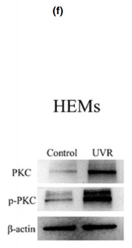
Application: WB Species: Human Sample: human epidermal melanocytes(HEMs)

Application: WB Species: rat Sample: HIP

Application: WB Species: Mice Sample: MC3T3-E1 cells
Application: WB Species: human Sample: CTC-TJH-01 cells
Application: WB Species: Human Sample: SH-SY5Y cells
Restrictive clause
Affinity Biosciences tests all products strictly. Citations are provided as a resource for additional applications that have not been validated by Affinity Biosciences. Please choose the appropriate format for each application and consult Materials and Methods sections for additional details about the use of any product in these publications.
For Research Use Only.
Not for use in diagnostic or therapeutic procedures. Not for resale. Not for distribution without written consent. Affinity Biosciences will not be held responsible for patent infringement or other violations that may occur with the use of our products. Affinity Biosciences, Affinity Biosciences Logo and all other trademarks are the property of Affinity Biosciences LTD.