.
Bands result from membrane strip incubation.")
.
Bands result from membrane strip incubation.")


 and mouse anti-beta tubulin Ab(T0023 1:200) for 1 hour at 37°C. An AlexaFluor594 conjugated goat anti-rabbit IgG(H+L) Ab(Red) and an AlexaFluor488 conjugated goat anti-mouse IgG(H+L) Ab(Green) were used as the secondary antibody.
The nuclear counter stain is DAPI(blue).")
: the relative expression of TGF-β1 (B), p-Smad2/3 (C) and BAMBI (D) were analyzed in the liver of TLR4wild/ TLR4mut mice infected with or without C. sinensis-infected by Western blotting. The blots of each group were run under the same experimental conditions and the images were from the same gel. The data were obtained from 3~5 mice of three-independent experiment. The values were expressed as mean±SEM.")
 of
immunolabeling for Fn and nuclear staining with DAPI. Scale bar, 50 ~μm.")
 and pCX43 (C) showed a significant decrease but TGF-β1 (D), Smad3 (E), and pSmad3 (F) showed a significant increase in the bladder detrusor after SCI. Those changes were more significant in transection than in hemisection of sacral spine cord. CX45 was not changed among three groups.")
 + TGF-β1 (0 ng/mL), corilagin (0 μM) + TGF-β1 (5
ng/mL) and corilagin (25 μM) + TGF-β1 (5 ng/mL) for 12 h. Smad2/3 is shown by green fluorescence and nuclei were stained with DAPI, which emits blue
fluorescence. Scale bars = 50 μm. D. Protein levels of Smad7 examined by western blot assay after HSFs were treated with corilagin for 3 days. GAPDH served as
control. n = 3. E. Protein levels of MMP2, MMP9, MMP13 and TIMP1 in HSFs after treatment with corilagin for 3 days. GAPDH served as control. n = 3. Data are
show as mean ± SD. *p < 0.05, **p < 0.01, ***p < 0.001. (For interpretation of the references to color in this figure legend, the reader is referred to the web version
of this article.)")
, corilagin (0 μM) + TGF-β1 (5
ng/mL) and corilagin (25 μM) + TGF-β1 (5 ng/mL) for 48 h. α-SMA is shown by green fluorescence and nuclei were stained with DAPI, which emits blue fluorescence.
Scale bars = 50 μm. B. qRT-PCR results of α-SMA mRNA levels in HSFs after incubation with TGF-β1 and corilagin for 3 days. n = 3. C. α-SMA protein levels detected
using western blot assay after treatment with TGF-β1 and corilagin for 3 days, and quantification results normalized to GAPDH. n = 3. Data are mean ± SD. *p <
0.05, ***p < 0.001. ns, no significance. (For interpretation of the references to color in this figure legend, the reader is referred to the web version of this article.)")

 Expression of proliferation- and migration-associated genes (PCNA, MMP9 and TIMP-1) were evaluated using western blotting in HEY cells. (C and D) Western blotting of proteins involved in integrin-β1-FAK signaling pathway in the KRT7-overexpressing HEY cells. (E) Expression of MMPs after knockdown of KRT7 in OVCAR433 cells. (F and G) Expression of the TGF-β signaling pathway-related proteins was evaluated by western blotting in KRT7-overexpressing HEY cells and KRT7-knockdown OVCAR433 cells. All experiments were performed at least three times. Results are presented as the mean ± standard deviation. **P<0.01. FAK, focal adhesion kinase; PCNA, proliferating cell nuclear antigen; FN, fibronectin; TIMP-1, TIMP metallopeptidase inhibitor 1; p-, phosphorylated; MMP, matrix metalloproteinase; KRT7, keratin 7; sh, short hairpin RNA; NC, negative control.")
, and western blot assay disclosed that the expressions of exosome markers CD9, CD63, CD81, TGF-β1 Flotillin-1 and EEF2 were elevated in Exosome group than those in NC group (sample from serum-free medium) (B).")
. Different letters marked above the bars are significantly different by an ANOVA multiple test (p < 0.05).")

, IL-6 (b) and IL-10 (c). Three samples per group were examined, and representative images are shown (Scale bar represents 50 µm).")
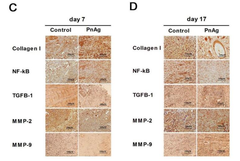
. Scale bar, 100 μm. b The expression of profibrotic proteins TGF-β1, fibronectin, collagen I, and collagen III in the kidney of unilateral urethral obstruction (UUO) mice, and quantitative analysis")
 Te expression levels of the renal fbrosis-associated genes TGF-β1, FN, Smad3, Col1a1, Col1a2, and α-SMA were quantitated in each of the groups. All groups were compared with OC. *stands for p<0.05, **stands for p<0.01, and ***stands for p<0.001. (b) Te western blot analysis of TGF-β1, Smad3, a-SMA, FN, Col1a1 and Col1a2.")

 Representative western blots and quantitative analysis of (B) TGF‑β1/β‑actin, (C) p‑Smad2/3/Smad2/3 and (D) p‑ERK1/2/ERK1/2 levels in rats.")
 Immunofluorescence was conducted to evaluate the expression of α-SMA (red). Nucleus was stained with DAPI
(blue). Scale bar: 50 μm. (b) Relative mRNA expression of Col1a1 and α-SMA. (c, d) Expressions of p-MEK1/2, MEK1/2, p-ERK1/2, ERK1/
2 were detected by western blot. GAPDH was conducted as a loading control. One-way ANOVA, **p < 0.01, ***p < 0.001,
****p < 0.0001.")


 reduces peritoneal
fibrosis (PF) relative protein and gene expression
in mice tested by western blotting and real-time
qPCR, respectively. The results of western blotting (A, B) and real-time qPCR (C) demonstrate
that, after intraperitoneal administration of
4.25% peritoneal dialysis solution, the levels of
α-SMA, Col-I, TGF-β1, SGLT-2 and p-Smad3 increase, and E-cadherin decreases in the submesothelial compact zone on day 28. Treatment
with Emp significantly reduces the levels of
α-SMA, Col-I, TGF-β1, and p-Smad3, and increases the levels of E-cadherin compared with
the effect of the peritoneal dialysis solution alone.
*P < 0.05, **P < 0.01, ***P < 0.001 versus PF
mice with peritoneal dialysis solution injected. N
= 6 per group.")
 TGF-β1 mRNA and protein levels were increased in HT22 cells in the presence of hemin. (C and D) HT22 cells were stimulated with different doses (0, 2, 5, and 10 ng/mL) of recombinant TGF-β1, and then assayed for SERPINE1 expression using qRT-PCR assay and Western blot. (E and F) HT22 cells were transfected with TGF-β1 siRNA, and then assayed for SERPINE1 expression using qRT-PCR assay and Western blot. **p <0.01 and ***p <0.001.")

 Western blot analysis for α-smooth muscle actin (α-SMA), collagen I, transforming growth factor (TGF)-β1 and connective tissue growth factor (CTGF). The relative expression of (B) α-SMA, (C) collagen I, (D) TGF-β1 and (E) CTGF. Data were shown as mean value ± SD (n=6). ***P<0.001, versus Control group. &&P<0.01, &&&P<0.001, versus NC agomir group.")
.
(A) The mRNA expression of TGF-β1. (B) The mRNA expression of SMAD2. (C) Representative images of the western blotting analysis for the quantification of TGF-β1, p-SMAD2, and SMAD2 in the heart tissues. (D) The protein expression of TGF-β1 in the heart tissues. (E) The protein expression of p-SMAD2/SMAD2 in the heart tissues. The data are expressed as the means ± SDs. ∗, P < 0.05 and ∗∗, P < 0.01 compared with the Sham group. #, P < 0.05 and ##, P < 0.01 compared with the AMI group. △, P < 0.05 and △△, P < 0.01 compared with the AMI group.")
. D, F, G. The expression levels of MMP2 (D, F) and MMP9 (D, G) in lung cancer tissues (n=6). D, H. The expression level of TGF-β1 in lung cancer tissues (n=6). D, I. The expression level of collagen type I in lung cancer tissues(n=6). Note: &, &&&& respectively represent a significant difference compared with the Saline group (P<0.05), (P<0.0001).")
. (A–F) Representative immunohistochemical staining of Collagen I, Fibronectin, PAI-1, TGF-β, IL-1β and IL-18 in the 4 groups (scale bar = 20 μm). (G–L) Quantitative analysis of the Collagen I, Fibronectin, PAI-1, TGF-β, IL-1β, and IL-18 protein expression shown in panels (A–F). *p < 0.05 versus the control group; #p < 0.05 versus the Ad-SC-shRNA group.")
 TGF-β1 protein levels. (B) α-SMA protein levels. Data are presented as mean ± SEM (n > 3 for each group). #P < 0.05 vs. CON, ∗P < 0.05 vs. DKD.")
, α-SMA, fibronectin, and collagen I expression in tracheal
scar tissues from normal, saline, and nintedanib (10 mg, 20 mg) treated rat groups. (A) TGF-β1 was detected in both the epithelial and submucosal layers (200); (B) α-SMA was detected in fibroblasts in submucosal layers (200); (C,D) fibronectin and collagen I were expressed in the
intercellular substance and submucosal cells in the stenotic tissues (200). Average optical density (AOD) date of four antibodies are presented as the means SEM. *P < .05, **P < .01, ***P < .001, ****P < .0001 compared with saline-treated group; ns = not significant. [Color
figure can be viewed in the online issue, which is available at www.laryngoscope.com.]")
 Relative mRNA level of IL10, Arg-1, and TGF-β1in the colon was measured through qPCR. Expression of IL-10, Arg-1, and TGF-β1 in colon tissue lysates were determined by Western blot from DSS-challenged mice. n = 3,.means ± SEM.*p < 0.05, **p < 0.01, ***p < 0.001.")
 CX3CR1 (red, stained with Alexa Fluor 594) and TGFβ1 (green, stained with Alexa Fluor 488) double labeling. TGFβ1 colocalized with CX3CR1 at 3 and 7 dpi, but not pre-injury or at 14 dpi. The asterisks indicate the lesion epicenter. Colocalization of the proteins is shown in yellow. Nuclear staining (DAPI) is shown in blue, and white arrowheads indicate the colocalization observed with a 40× objective. Scale bars: 200 μm in the left three columns; 20 μm in the right column. (B) Percentage of TGFβ1+CX3CR1+ cells relative to the total number of CX3CR1+ cells in the injured spinal cord. The data are presented as the mean ± SEM (n = 3 independent experiments). ****P < 0.0001 (one-way analysis of variance followed by Tukey's post hoc test). CX3CR1: C-X3-C motif chemokine receptor 1; DAPI: 4′,6-diamidino-2-phenylindole, dihydrochloride; dpi: days post-injury; ND: not determined; TGFβ1: transforming growth factor-β1.")
 Western blotting shows the expression of M1 (iNOS) or M2 (Arg1) polarization markers and TGFβ1 in BV-2 cells after polarization induction. (B) Quantitative analysis of iNOS expression shown in (A). β-Actin was used as the loading control. iNOS was expressed at significantly higher levels in M1-type microglia than in M0- or M2-type microglia. (C) Quantitative analysis of Arg1 protein expression shown in (A). Arg1 was expressed at significantly higher levels in M2-type microglia than in M0- or M1-type microglia. (D) Quantitative analysis of TGFβ1 protein expression shown in (A). TGFβ1 was expressed at significantly higher levels in M1-type microglia than in M0- or M2-type microglia. The data are presented as the mean ± SEM (n = 4) and were analyzed by one-way analysis of variance, followed by Tukey's post hoc test. ***P < 0.001, ****P < 0.0001. (E) Representative immunocytochemistry images of TGFβ1 (green, stained with Alexa Fluor 488) in BV-2 cells after polarization induction. The TGFβ1 fluorescence intensity was strongest in M1-type microglia, suggesting that these cells expressed the highest levels of TGFβ1. DAPI (blue) was used to stain the nuclei. At least three independent replicates were performed for each experiment. Scale bar: 50 μm. Arg1: Arginine 1; DAPI: 4′,6-diamidino-2-phenylindole, dihydrochloride; iNOS: inducible nitric oxide synthase; TGFβ1: transforming growth factor-β1.")

 Representative results of Western blot analysis of α-SMA,
Collagen I and TGF-β1 in rats from all five groups. (B) Expression of α-SMA in all five groups with β-actin as the loading control is presented
as a bar graph. (C) Expression of Collagen I in all five groups with β-actin as the loading control is presented as a bar graph. (D) Expression
of TGF-β1 in all five groups with β-actin as the loading control is presented as a bar graph. (E) Representative images showing smooth
muscle and collagen staining in all five groups. Smooth muscle and collagen in the corpus cavernosum are stained red and blue, respectively.
Original magnification, 50× and 200×. (F) Representative results of immunohistochemistry for TGF-β1 in all five groups (50× and 200×). The
arrows indicate TGF-β1 expression. Data are expressed as the mean ± standard deviation. &P < .05 compared with the Con group. #
P < .05
compared with the DMED group. *
P < .05 compared with the DMED + CCSMC-EXO group. $
P < .05 compared with the DMED + BMSCEXO group. CCSMC-EXOs: exosomes derived from corpus cavernosum smooth muscle cells; BMSC-EXOs: exosomes derived from bone
marrow stem cells; ADSC-EXOs: exosomes derived from adipose-derived stem cells; DMED: diabetes mellitus-induced erectile dysfunction")
 Representative images showing the distribution of PAI-1 in cardiac tissues after immunofluorescence staining. Scar bar = 50 μm. (b, c) Relative mRNA and protein expression of urokinase plasminogen activator (uPA) and tissue-type plasminogen activator (tPA) in the cardiac tissues. (d) Western blot analysis for TGF-β1R, TGF-β1, p-Smad2/3, and Smad2/3. Relative protein expression of (e) PAI-1, (f) TGF-β1R, (g) TGF-β1, (h) p-Smad2/3, and (i) Smad2/3 in the cardiac tissues. Values are expressed as mean ± SD (n = 6). +++p < 0.001vs. sham group. ∗p < 0.05, ∗∗p < 0.01, ∗∗∗p < 0.001vs. MI/R group.")
 The representative band of Western blot. (b)–(h) The quantitative result of Western blot. Data were expressed as mean ± SD. #P < 0.05/##p < 0.01 vs. control; ∗p < 0.05/∗∗p < 0.01 vs. the BLM group. n = 3.")
 mHSCs were co-cultured with the CMs from RAW264.7 cells with LPS/IFNγ and PHI treatment for 12 h. (b-g) The expression of MMP2, TIMP1, TGF-β, α-SMA, COL1 and NF-κB mRNA in mHSCs after co-culture with macrophage-derived CMs for 24 h was detected by RT-qPCR (n = 3). (h) The expression of TGF-β, α-SMA, COL1, P65 and P-P65 proteins in mHSCs after co-culture with macrophage-derived CMs for 24 h was detected by western blotting. (i) The relative quantification of TGF-β, α-SMA, COL1 and P-P65/P65 protein expression in western blotting results was analyzed by ImageJ software (n = 3). (j) The expression of cytoplasm and nucleus NF-κB P65 proteins in mHSCs after co-culture with macrophage-derived CMs for 24 h was detected by western blotting. (k, l) The relative quantification of cytoplasm and nucleus NF-κB P65 protein expression in western blotting results was analyzed by ImageJ software (n = 3). (m) Immunofluorescence staining of α-SMA in mHSCs after co-culture with CMs from RAW264.7 cells with different treatments for 24 h (Scale bar=100 µm). Results are presented as mean ± SD. ###P")

. Western blotting analysis of NF-κB, TGF-β, PXR, IL-6, α-SMA in livers, and the datas were normalized to that of GAPDH (A–H), Western blotting analysis of Bax, and PXR in HepG2 cells, and the datas were normalized to that of GAPDH (H,I). Values are the mean ± standard deviation.")
. NC, normal control group; BLM, bleomycin-induced systemic sclerosis model group; PESV-L, low-dose PESV intervention group; PESV-M, medium-dose PESV intervention group; PESV-H, high-dose PESV intervention group; DXM, dexamethasone intervention group.")
 Immunohistochemical detection of arginase-1, (b) Statistical expressions of arginase-1 by an immunohistochemical assay (p")
 Expression levels of TGFb1, SMAD2/3, and quantification. (d‒g) Expression levels of TGFb2, TGFb3, pSMAD2/3 (Thr8), and quantification. (h‒k) Expression levels of Akt and pAkt and quantification. n ¼ 3 biological replicates. Data are represented as mean SD; one-way ANOVA with Fisher’s posthoc test. *P < 0.05 versus control and #P < 0.05 versus FB-Exo. Akt, protein kinase B; ESC-Exo, epidermal stem cellederived exosome; FB, fibroblast; FB-Exo, fibroblast-derived exosome; pAkt, phosphorylated protein kinase B; pSMAD, phosphorylated SMAD.")
. (A) Masson’s trichrome staining of uterine horns (×400). (B) Representative images of IHC for the fibrosis associated marker COL I (×400). (C) Representative images of IHC for the pro-fibrogenic mediator TGF-β 1 (×400). Scale bar=500 µm. (D) Ratio of the area with fibrosis to the total endometrial area in each group. (E) Quantification of COL I-positive expression. (F) Quantification of TGF-β1-positive expression. All data are expressed as the mean ± SEM (*P")
. As compared to the normal group, the smad3 and TGF-β1 proteins elevated in all the groups excepting the high dose of CA-treated group(P")
. As compared to the normal group, the smad3 and TGF-β1 proteins elevated in all the groups excepting the high dose of CA-treated group(P")
 and (B) Transmission electron microscopy of the enriched extracellular vesicles (EVs) isolated from the sera of E. multilocularis-infected (A) and uninfected mice (B). (C) The diameter distribution of the purified EVs. (D) Western blotting analysis of mammalian EV-marker CD63 in the purified EVs. (E) Heatmap of differentially abundance proteins between the serum exosomes from E. multilocularis-infected and uninfected mice. (F) Western blotting analysis the abundance of TGF-β1, GPATC8, LRP1, and AMY1 during E. multilocularis infection. https://doi.org/10.1371/journal.pntd.0010814.g001")
 of RIPK1, RIPK3, MLKL (necroptosis core mediators), NRGPI oncogenes (SERPINE1, GPX3, GRP, FCN1, CYTL1, CNTN1, PLCL1, and APOD), markers of WNT signaling pathway (WNT2B, WNT9A), TGF-β signaling pathway (TGFB1, TGFB3), and macrophage (CD63, CD206, CD163) in the clinical samples of stomach adenocarcinoma.")
 and high-dose aspirin (300 mg/kg + CCl4). ns, not significant. *P")
 macrophages. (A) Different morphologic appearances of THP-1 cells that were untreated (THP-1), treated with PMA (M0) or treated with PMA and subsequently with IL4/IL13 (M2). THP-1 cells grew in suspension, while M0 and M2 were larger with increased adherence and more granules in the cytoplasm. Scale bar: 50 μm. (B) Representative flow cytometric analysis of untreated THP-1, M0 and M2 cells stained with allophycocyanin-conjugated monoclonal antibody anti-CD206. The MFI is shown on the x-axis, and the relative cell number on the y-axis. (C) Representative immunofluorescence images showing increased expression of CD206 in M2 cells compared to M0 macrophages. Scale bars: 10 μm. (D) Quantification of the results in (C). **P")
 Representative images of Periodic Acid Schiff staining (PAS, ×400, bar: 20 μm). (B) Representative images of Masson's trichrome staining (Masson, ×400, bar: 20 μm). (C) Statistics of mesangial area. (D) Statistics of mesangial density. (E) Statistical result of Masson. (F) Representative WB images of TGF-β1, Smad2/3, and p-Smad2/3. (G) Statistical results of TGF-β1. (H) Statistical results of p-Smad2/3 to Smad2/3. Data are expressed as mean ± SD, n = 4. **P < 0.01 versus Control; #P < 0.05, ##P < 0.01 versus Model.")
. All experiments were repeated three times.")
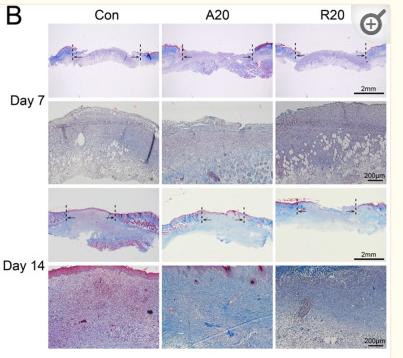
 Representative micrographs showing collagen deposition in obstructed kidneys. Mice were pretreated with Apcin (5 mg/kg per day) by intraperitoneal injection for 1 day. UUO surgery was then performed, and Apcin was administered continuously for the next 7 days until mice were sacrificed on day 7. Paraffin sections were subjected to Masson's trichrome staining for collagen deposition. Scale bar: 50 μm. (B) Graphic presentation showing the fibrotic area in kidney tissues among groups as indicated (n = 8 in vehicle Sham group; n = 7 in Apcin Sham group; n = 7 in vehicle UUO group; n = 7 in Apcin UUO group). The quantification of fibrotic area was performed by Image-Pro Plus. (C) Representative micrographs of Sirius red staining in mice after UUO. Scale bar: 50 μm. (D and E) Representative micrographs and quantitative data for FN1 immune staining in kidney tissues (n = 4). Scale bar: 50 μm. (F and G) Representative micrographs and quantitative data showing α-SMA expression in kidney tissues among groups as indicated (n = 4). Scale bar: 50 μm. Positive signal quantification was performed using Image-Pro Plus. (H and I) The mRNA level of Collagen I and Collagen III were evaluated by qRT-PCR (n = 8 in vehicle Sham group; n = 7 in Apcin Sham group; n = 7 in vehicle UUO group; n = 7 in Apcin UUO group). (J and K) Representative Western blot and quantitative data showing renal expression of Collagen I, Collagen III, TGF-β1 and CDC20 (n = 3 in sham group; n = 4 in UUO group). The values represent means ± SEM. P values were shown in figure.")
 Immunohistochemical staining of COL1 (400×, scale bar = 40 µm). (B) The mean density of COL1 staining. (C) The mRNA expression of COL1. (D) COL1, α-SMA and GAPDH were detected by WB. (E and F) Relative expressions of COL1 and α-SMA. (G) Samd2/3, p-Samd2/3, TGF-β and GAPDH were detected by WB. (H and I) Relative expressions of p-Samd2/3/Samd2/3 and TGF-β. (J) Relative expression of TGF-β mRNA. Data are expressed as mean ± SD, q-PCR n = 3, others n = 4. **P < 0.01 as compared with Control group. #P < 0.05, ##P < 0.01 as compared with HFD + STZ group.")
. #p")
, P < 0.05). The effect of ASP on the TGF-β/Smad4 pathway was further evaluated. TGF-β and Smad4 protein expressions were both dose-dependently decreased by ASP treatment, as shown by western blotting (Figure 2(b), P < 0.05).")
 Cell viability was evaluated by the CCK-8 assay. (B) The qRT-PCR analysis of TGFβ1, COL1A2, and ACTA2 (α-SMA) mRNA levels of LF cells which were treated with the optimal concentration of the THBS1 protein. (C) Western blotting assay showing protein expression levels of TGFβ1, Smad3, COL1A2, and α-SMA of LF cells which were treated with various dosages of THBS1 for 24 h. (D) Immunohistochemical staining of p-Smad3 of the LF samples from LFH group (n = 6) and non-LFH group (n = 6), and the percentage of positive cells was quantified. Scale bar = 20 μm. (E) Protein levels of TGFβ1, Smad3, COL1A2, and α-SMA of LF cells with different treatments. (F) Immunofluorescence staining of p-Smad3 of LF cells with different treatments. Nuclei were stained with DAPI (blue). p-Smad3 was stained red. Scale bar = 20 μm. (G) Double-labeling immunohistofluorescence staining of TGFβ1 and COL1A2 of cells with different treatments. Nuclei were stained with DAPI (blue). TGFβ1 was stained green. COL1A2 was stained red. Scale bar = 20 μm. These cells were mechanically stretched for 16 h. THBS1 and LSKL stimulated cells for 24 h. These data are representative of three independent experiments. Error bars: mean ± S.D. ns, not significant. ***p < 0.001.")
 The thickness of LF was measured by MRI before operation. The thickness of LF at the L4/5 facet joint level was indicated by red arrow. (B) Representative tissues of LF acquired from patients with spinal surgery. The top three were from non-LFH patients and the bottom three were from LFH patients. Scale bar = 5 mm. (C) Representative images of H&E, MASSON, and EVG staining of the non-LFH group (n = 6) and the LFH group (n = 6). The yellow dotted area represents collagen fibers. Scale bar = 20 μm. (D) Immunohistochemical staining of THBS1, TGFβ1, COL1A2, and α-SMA in the LF samples from LFH group (n = 6) and non-LFH group (n = 6), and the percentage of positive cells was quantified. Scale bar = 20 μm. (E) TEM of LF ultrastructure of non-LFH and LFH groups. The yellow area represents elastin fibers. Scale bar = 8 μm. (F) The qRT-PCR analysis results showing that LFH group (n = 6) had a higher mRNA level of THBS1, TGFβ1, ACTA2 (α-SMA), and COL1A2 compared with non-LFH group (n = 6). The 2−ΔΔCt method was used to normalized with GAPDH. (G) Western blotting assay of THBS1, TGFβ1, COL1A2, and α-SMA protein levels of LF samples of the two groups. GAPDH served as the loading control. EVG: verhoeff's van gieson, TEM: transmission electron microscope, EF: elastic fiber, CF: collagen fiber. **p < 0.01, ***p < 0.001.")
, α-SMA (B), Erbb4-IR (C) and miR-29b (D) in different groups was detected by qRT-PCR (n = 6), the results were presented as mean ± SD, *P")
 Down-regulation of GLI2 is accompanied by down-regulation of PD-L1, TGF-beta, IL6 and snail1. (E–H) Up-regulation of GLI2 is accompanied with increased expression of PD-L1, TGF-beta, and IL6. (I) Co-culture process. (J–L) Down-regulation of tumor-derived GLI2 decreases tumor-derived TGF-beta, PDL1 and IL6, likewise the NK-derived TIM-3 and PD1, while NK-derived IFN-gamma is increased. TGF-beta active protein powder restores the expression of tumor-derived PDL1 and IL6, and NK-derived TIM-3 and PD1. (M) Down-regulation of tumor-derived GIL2 leads increased secretion of NK-derived IFN-gamma, detected by Elisa.")
 Post-treatment results of fecal occult blood tests for each group. (B) Post-treatment comparison of body weights among the different groups of mice. (C) Post-treatment comparison of DAI scores across the various groups of mice. (D) Appearance and morphology of colon in each group of mice. (E) HE staining of colon tissue in each group of mice. The microscope has a magnification power of 200X, scale bar 100 μm. (G–J) Protein expression levels of IL-1β, IL-4, IL-10, and TGF-β1 in colon tissue of each group of mice. N = 10 mice for each group. Compared with the CON group, △p")
. C Measurement of TNF-alpha concentration in serum using the Mouse TNF-alpha ELISA Kit on days 28 and 56. D Changes in HYP content in lung tissues of each group on days 28 and 56. Data are presented as mean ± SD, n = 5 per group. (C: control group; M: model group; E: exosome group; H: stem cell group. *P")
 and TGF-β1 (B) expression in renal tissues of mice treated with different doses of Ang-(1–7): low (144 µg/kg), medium (288 µg/kg), or high (576 µg/kg), for 1 or 7 days (400 × magnification). Data were represented as mean ± SD (n = 3). **P")
 Masson’s trichrome staining and HE is staining of peritoneal tissue sections (×400). (b) Immunohistochemical staining of SOCS1, α-SMA, and Collagen I in peritoneal tissue sections (×400). (c) Expression levels of TGF-β, Smad2/3, p-Smad2/3, and SOCS1 were detected using protein blotting. Each group contained eight rats, and three fields of view were randomly selected for each group. The percentage of the stained area was calculated for statistical analysis. All immunoblotting experiments were performed independently and repeated at least three times. Values are expressed as mean ± standard deviation. *p < 0.05 vs. Control; **p < 0.01 vs. Control; ***p < 0.001 vs. Control.")
 and Smad3 (right) in Normal/Cancer, as analyzed using bioinformatics platforms (GEPIA and XIANTAO). *P")
. ***P")
. B Intergroup comparison of α-SMA integrated optical density (IOD) (n = 6). ***P")
 Western blot analysis of TGF-β1, TGF-βR1, p-Smad2, Smad2, p-Smad3, and Smad3 protein levels (fold change normalized to GAPDH or total Smad2/3) in lung tissues from sham, COPD, and COPD + OMT mice. (F) Serum TGF-β1 concentration (ng/mL) quantified by ELISA. N = 8 mice/group. (G) TGF-β1 secretion (ng/mL) in HBE cell supernatants after 10% CSE (v/v, 48 h) ± OMT (80 μM, 24-h pretreatment). (H–I) Time-dependent phosphorylation of Smad2/3 (fold change normalized to total Smad2/3) in HBE cells exposed to 10% CSE (v/v, 0–60 min) ± OMT (80 μM, 24-h pretreatment). (J–L) Immunofluorescence staining (scale bar: 50 μm) and quantification of nuclear Smad2/3 intensity (RFU) in HBE cells treated with 10% CSE (v/v, 30 min) ± OMT (80 μM, 24-h pretreatment). N = 3. ###p")
 Representative images of protein bands. (b-e) TGF-β1, (c) p-ERK1/2 / ERK1/2, (d) p-Smad2/Smad2, (e) pSmad3/Smad3, (f) type II collagen, (g) ALP, (h) TRACP, and (i) BMP7 were measured by western blot. (n=3, means ± SD) **P")
. D Changes in protein ratios of iNOS and Arg1 (n = 6 independent experiments). E-H Western blot detects changes in protein levels of anti-inflammatory cytokines IL-4, IL-10, and TGF-β in mouse brain (n = 6 independent experiments). I-K Western blot detects changes in protein levels of anti-inflammatory cytokines IL-1β and IFN-γ in mouse substantia nigra (n = 6 independent experiments). L-R Western blot detected the protein levels of RANK, RANKL, OPG, p-NIK, NIK, p-IKK, IKK, p-NF-κB2, and NF-κB2 protein level changes (n = 6 independent experiments). iNOS is a marker for neurotoxic microglial, Arg1 (arginase-1) is a marker for neuroprotective microglial. Data are mean ± SEM.")
 CCK-8 assay was used to assess the cell viability of HaCaT and fibroblast cells after treatment with Elesclomol+Cu2+ (1 μM) across different concentration gradients (A) and time gradients (B). (C and D) CCK-8 assay was performed to evaluate the cell viability of Hacat and fibroblast cells treated with different cell death inhibitors (TTM, Nec-1, Fer-1, CQ, ZVF). (E) IHC to verify the expression of ACO2 in normal tissues and DFU tissues, Scale bars = 100 μm (F) Western blotting confirmed the change of ACO2 protein levels in Hacat and fibroblast. (G and H) qRT-PCR confirmed the knockdown of TMPO in Hacat and fibroblast cells by shRNA on mRNA levels. (I and J) Western blotting confirmed the knockdown of TMPO in Hacat and fibroblast cells by shRNA on protein levels. (K and M) Cell growth curve of Hacat and fibroblast cells transfected with Scramble or shTMPO. (L, N, O, P) Wound healing assays of Hacat and fibroblast cells transfected with Scramble or shTMPO. (Q and R) The protein levels of TGF-β1, Smad2/3, p-Smad2/3, and P21 in Hacat and fibroblast cells transfected with Scramble or shTMPO. n = 3, data were represented as mean ± SEM.")
 Correlation analysis of intestinal flora and bile acid metabolism. (B) FOXP3 and TGF-β1 protein expressions (control images are re-used for illustrative purposes, n = 3). (C) mRNA expression of FOXP3 and TGF-β1 (compared with the control: #p")
| Product: | TGF beta 1 Antibody |
| Catalog: | AF1027 |
| Description: | Rabbit polyclonal antibody to TGF beta 1 |
| Application: | WB IHC IF/ICC |
| Cited expt.: | WB, IHC, IF/ICC |
| Reactivity: | Human, Mouse, Rat |
| Prediction: | Pig, Bovine, Horse, Sheep, Dog |
| Mol.Wt.: | 44~65kd(precursor), 28kD(dimer), 15kD(monomer)(Observed); 44kD(Calculated). |
| Uniprot: | P01137 |
| RRID: | AB_2835389 |
Control Products

Related Downloads
Protocols
Product Info
*The optimal dilutions should be determined by the end user. For optimal experimental results, antibody reuse is not recommended.
*Tips:
WB: For western blot detection of denatured protein samples. IHC: For immunohistochemical detection of paraffin sections (IHC-p) or frozen sections (IHC-f) of tissue samples. IF/ICC: For immunofluorescence detection of cell samples. ELISA(peptide): For ELISA detection of antigenic peptide.
Cite Format: Affinity Biosciences Cat# AF1027, RRID:AB_2835389.
Fold/Unfold
Cartilage-inducing factor; CED; Differentiation inhibiting factor; DPD1; LAP; Latency-associated peptide; Prepro transforming growth factor beta 1; TGF beta 1; TGF beta; TGF beta 1 protein; TGF-beta 1 protein; TGF-beta-1; TGF-beta-5; TGF-beta1; TGFB; Tgfb-1; tgfb1; TGFB1_HUMAN; TGFbeta; TGFbeta1; Transforming Growth Factor b1; Transforming Growth Factor beta 1; Transforming growth factor beta 1a; transforming growth factor beta-1; transforming growth factor, beta 1; Transforming Growth Factor-ß1;
Immunogens
A synthesized peptide derived from human TGF beta1, corresponding to a region within C-terminal amino acids.
Highly expressed in bone (PubMed:11746498, PubMed:17827158). Abundantly expressed in articular cartilage and chondrocytes and is increased in osteoarthritis (OA) (PubMed:11746498, PubMed:17827158). Colocalizes with ASPN in chondrocytes within OA lesions of articular cartilage (PubMed:17827158).
- P01137 TGFB1_HUMAN:
- Protein BLAST With
- NCBI/
- ExPASy/
- Uniprot
MPPSGLRLLLLLLPLLWLLVLTPGRPAAGLSTCKTIDMELVKRKRIEAIRGQILSKLRLASPPSQGEVPPGPLPEAVLALYNSTRDRVAGESAEPEPEPEADYYAKEVTRVLMVETHNEIYDKFKQSTHSIYMFFNTSELREAVPEPVLLSRAELRLLRLKLKVEQHVELYQKYSNNSWRYLSNRLLAPSDSPEWLSFDVTGVVRQWLSRGGEIEGFRLSAHCSCDSRDNTLQVDINGFTTGRRGDLATIHGMNRPFLLLMATPLERAQHLQSSRHRRALDTNYCFSSTEKNCCVRQLYIDFRKDLGWKWIHEPKGYHANFCLGPCPYIWSLDTQYSKVLALYNQHNPGASAAPCCVPQALEPLPIVYYVGRKPKVEQLSNMIVRSCKCS
Predictions
Score>80(red) has high confidence and is suggested to be used for WB detection. *The prediction model is mainly based on the alignment of immunogen sequences, the results are for reference only, not as the basis of quality assurance.
High(score>80) Medium(80>score>50) Low(score<50) No confidence
Research Backgrounds
Transforming growth factor beta-1 proprotein: Precursor of the Latency-associated peptide (LAP) and Transforming growth factor beta-1 (TGF-beta-1) chains, which constitute the regulatory and active subunit of TGF-beta-1, respectively.
Required to maintain the Transforming growth factor beta-1 (TGF-beta-1) chain in a latent state during storage in extracellular matrix. Associates non-covalently with TGF-beta-1 and regulates its activation via interaction with 'milieu molecules', such as LTBP1, LRRC32/GARP and LRRC33/NRROS, that control activation of TGF-beta-1. Interaction with LRRC33/NRROS regulates activation of TGF-beta-1 in macrophages and microglia (Probable). Interaction with LRRC32/GARP controls activation of TGF-beta-1 on the surface of activated regulatory T-cells (Tregs). Interaction with integrins (ITGAV:ITGB6 or ITGAV:ITGB8) results in distortion of the Latency-associated peptide chain and subsequent release of the active TGF-beta-1.
Transforming growth factor beta-1: Multifunctional protein that regulates the growth and differentiation of various cell types and is involved in various processes, such as normal development, immune function, microglia function and responses to neurodegeneration (By similarity). Activation into mature form follows different steps: following cleavage of the proprotein in the Golgi apparatus, Latency-associated peptide (LAP) and Transforming growth factor beta-1 (TGF-beta-1) chains remain non-covalently linked rendering TGF-beta-1 inactive during storage in extracellular matrix. At the same time, LAP chain interacts with 'milieu molecules', such as LTBP1, LRRC32/GARP and LRRC33/NRROS that control activation of TGF-beta-1 and maintain it in a latent state during storage in extracellular milieus. TGF-beta-1 is released from LAP by integrins (ITGAV:ITGB6 or ITGAV:ITGB8): integrin-binding to LAP stabilizes an alternative conformation of the LAP bowtie tail and results in distortion of the LAP chain and subsequent release of the active TGF-beta-1. Once activated following release of LAP, TGF-beta-1 acts by binding to TGF-beta receptors (TGFBR1 and TGFBR2), which transduce signal. While expressed by many cells types, TGF-beta-1 only has a very localized range of action within cell environment thanks to fine regulation of its activation by Latency-associated peptide chain (LAP) and 'milieu molecules' (By similarity). Plays an important role in bone remodeling: acts as a potent stimulator of osteoblastic bone formation, causing chemotaxis, proliferation and differentiation in committed osteoblasts (By similarity). Can promote either T-helper 17 cells (Th17) or regulatory T-cells (Treg) lineage differentiation in a concentration-dependent manner (By similarity). At high concentrations, leads to FOXP3-mediated suppression of RORC and down-regulation of IL-17 expression, favoring Treg cell development (By similarity). At low concentrations in concert with IL-6 and IL-21, leads to expression of the IL-17 and IL-23 receptors, favoring differentiation to Th17 cells (By similarity). Stimulates sustained production of collagen through the activation of CREB3L1 by regulated intramembrane proteolysis (RIP). Mediates SMAD2/3 activation by inducing its phosphorylation and subsequent translocation to the nucleus. Can induce epithelial-to-mesenchymal transition (EMT) and cell migration in various cell types.
Transforming growth factor beta-1 proprotein: The precursor proprotein is cleaved in the Golgi apparatus by FURIN to form Transforming growth factor beta-1 (TGF-beta-1) and Latency-associated peptide (LAP) chains, which remain non-covalently linked, rendering TGF-beta-1 inactive.
N-glycosylated. Deglycosylation leads to activation of Transforming growth factor beta-1 (TGF-beta-1); mechanisms triggering deglycosylation-driven activation of TGF-beta-1 are however unclear.
Secreted>Extracellular space>Extracellular matrix.
Secreted.
Highly expressed in bone. Abundantly expressed in articular cartilage and chondrocytes and is increased in osteoarthritis (OA). Colocalizes with ASPN in chondrocytes within OA lesions of articular cartilage.
The 'straitjacket' and 'arm' domains encircle the Transforming growth factor beta-1 (TGF-beta-1) monomers and are fastened together by strong bonding between Lys-56 and Tyr-103/Tyr-104.
The cell attachment site motif mediates binding to integrins (ITGAV:ITGB6 or ITGAV:ITGB8) (PubMed:28117447). The motif locates to a long loop in the arm domain called the bowtie tail (PubMed:28117447). Integrin-binding stabilizes an alternative conformation of the bowtie tail (PubMed:28117447). Activation by integrin requires force application by the actin cytoskeleton, which is resisted by the 'milieu molecules' (such as LTBP1, LRRC32/GARP and/or LRRC33/NRROS), resulting in distortion of the prodomain and release of the active TGF-beta-1 (PubMed:28117447).
Belongs to the TGF-beta family.
Research Fields
· Cellular Processes > Cell growth and death > Cell cycle. (View pathway)
· Cellular Processes > Cell growth and death > Cellular senescence. (View pathway)
· Environmental Information Processing > Signal transduction > MAPK signaling pathway. (View pathway)
· Environmental Information Processing > Signaling molecules and interaction > Cytokine-cytokine receptor interaction. (View pathway)
· Environmental Information Processing > Signal transduction > FoxO signaling pathway. (View pathway)
· Environmental Information Processing > Signal transduction > TGF-beta signaling pathway. (View pathway)
· Environmental Information Processing > Signal transduction > Hippo signaling pathway. (View pathway)
· Human Diseases > Endocrine and metabolic diseases > Non-alcoholic fatty liver disease (NAFLD).
· Human Diseases > Infectious diseases: Parasitic > Leishmaniasis.
· Human Diseases > Infectious diseases: Parasitic > Chagas disease (American trypanosomiasis).
· Human Diseases > Infectious diseases: Parasitic > Malaria.
· Human Diseases > Infectious diseases: Parasitic > Toxoplasmosis.
· Human Diseases > Infectious diseases: Parasitic > Amoebiasis.
· Human Diseases > Infectious diseases: Bacterial > Tuberculosis.
· Human Diseases > Infectious diseases: Viral > Hepatitis B.
· Human Diseases > Infectious diseases: Viral > HTLV-I infection.
· Human Diseases > Cancers: Overview > Pathways in cancer. (View pathway)
· Human Diseases > Cancers: Overview > Proteoglycans in cancer.
· Human Diseases > Cancers: Specific types > Colorectal cancer. (View pathway)
· Human Diseases > Cancers: Specific types > Renal cell carcinoma. (View pathway)
· Human Diseases > Cancers: Specific types > Pancreatic cancer. (View pathway)
· Human Diseases > Cancers: Specific types > Chronic myeloid leukemia. (View pathway)
· Human Diseases > Cancers: Specific types > Hepatocellular carcinoma. (View pathway)
· Human Diseases > Cancers: Specific types > Gastric cancer. (View pathway)
· Human Diseases > Immune diseases > Inflammatory bowel disease (IBD).
· Human Diseases > Immune diseases > Rheumatoid arthritis.
· Human Diseases > Cardiovascular diseases > Hypertrophic cardiomyopathy (HCM).
· Human Diseases > Cardiovascular diseases > Dilated cardiomyopathy (DCM).
· Organismal Systems > Development > Osteoclast differentiation. (View pathway)
· Organismal Systems > Immune system > Th17 cell differentiation. (View pathway)
· Organismal Systems > Immune system > Intestinal immune network for IgA production. (View pathway)
· Organismal Systems > Endocrine system > Relaxin signaling pathway.
References

Application: WB Species: human Sample:
Application: IHC Species: rat Sample:
Application: IHC Species: Mice Sample: skin

Application: WB Species: Human Sample: HK2 cells
Restrictive clause
Affinity Biosciences tests all products strictly. Citations are provided as a resource for additional applications that have not been validated by Affinity Biosciences. Please choose the appropriate format for each application and consult Materials and Methods sections for additional details about the use of any product in these publications.
For Research Use Only.
Not for use in diagnostic or therapeutic procedures. Not for resale. Not for distribution without written consent. Affinity Biosciences will not be held responsible for patent infringement or other violations that may occur with the use of our products. Affinity Biosciences, Affinity Biosciences Logo and all other trademarks are the property of Affinity Biosciences LTD.